. 2010 Nov 29;191(5):933-42.
doi: 10.1083/jcb.201008084.

Mitochondrial membrane potential regulates PINK1 import and proteolytic destabilization by PARL

Affiliations

Mitochondrial membrane potential regulates PINK1 import and proteolytic destabilization by PARL

Seok Min Jin et al. J Cell Biol. .

Abstract

PINK1 is a mitochondrial kinase mutated in some familial cases of Parkinson's disease. It has been found to work in the same pathway as the E3 ligase Parkin in the maintenance of flight muscles and dopaminergic neurons in Drosophila melanogaster and to recruit cytosolic Parkin to mitochondria to mediate mitophagy in mammalian cells. Although PINK1 has a predicted mitochondrial import sequence, its cellular and submitochondrial localization remains unclear in part because it is rapidly degraded. In this study, we report that the mitochondrial inner membrane rhomboid protease presenilin-associated rhomboid-like protein (PARL) mediates cleavage of PINK1 dependent on mitochondrial membrane potential. In the absence of PARL, the constitutive degradation of PINK1 is inhibited, stabilizing a 60-kD form inside mitochondria. When mitochondrial membrane potential is dissipated, PINK1 accumulates as a 63-kD full-length form on the outer mitochondrial membrane, where it can recruit Parkin to impaired mitochondria. Thus, differential localization to the inner and outer mitochondrial membranes appears to regulate PINK1 stability and function.


Figures

Figure 1.
Figure 1.
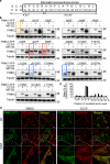
Constitutive cleavage of PINK1 is mediated by PARL. (a) HeLa cells were transfected with scrambled control siRNA or PARL siRNA. After 4 h incubation with or without 10 µM CCCP, mitochondria were isolated, and mitochondrial protein extracts were assayed for endogenous levels of PINK1 and PARL by immunoblotting. VDAC1 is a mitochondrial marker. (b) MEFs from PARL WT and KO mice were transfected with PINK1-V5/His for 2 d and treated with DMSO or 10 µM CCCP for 4 h. Exogenous PINK1 levels were assayed by immunoblotting. Tubulin is a loading control. (c) PARL WT and KO MEFs were transfected with PINK1-V5/His as in b and treated with DMSO or 10 µM MG132. After 4 h of treatment, cells were fractionated, and exogenous PINK1 levels in mitochondrial fraction were measured with immunoblotting. VDAC1, mitochondrial loading control. (d) [35S]methionine-labeled PINK1 was incubated for different times with mitochondria isolated from PARL WT or KO MEFs in the presence or absence of 1 µM CCCP. After import, samples were treated with or without 5 µg/ml PK. Radiolabeled PINK1 was detected using digital autoradiography. Asterisks, nonspecific bands. (e) [35S]-PINK1 was imported into PARL KO mitochondria for 60 min as in d, and these mitochondria were incubated in the presence or absence of high PK (100 µg/ml) for 10 min. Hsp70, Htra2/Omi, and Tom20 were identified by immunoblotting as markers for mitochondrial matrix, inter membrane space, and outer membrane, respectively. (f) HeLa cells stably expressing YFP-Parkin were transfected with control (siCtrl) or PARL siRNA (siPARL) for 192 h. After transfection, cells were treated with either DMSO or 10 µM CCCP for 1 h, stained with TMRE, and analyzed by live cell imaging. Bars, 20 µm. (g) PARL WT and KO MEFs transfected with PINK1-YFP and mCherry-Parkin were treated with 10 µM DMSO or CCCP for 3 h. Cells (≥50/treatment) were counted for mitochondrial translocation of Parkin. Counting results were represented as mean ± SEM from four replicates. Blue arrowheads, FL PINK1; orange arrowheads, ΔMTS-PINK1; red arrowheads, 52-kD PINK1.
Figure 2.
Figure 2.
The 52-kD form of endogenous PINK1 is found inside mitochondria and does not recruit Parkin. (a) HeLa cells were initially treated with 50 µM MG132 for 10 h and then together with 10 µM CCCP for a final 3 h. Cells were fractionated and analyzed by immunoblotting using antibodies against the indicated proteins. (b) The mitochondrial fraction from panel a was subjected to alkaline extraction using sodium carbonate and immunoblotted for PINK1, Cyt c, and Tim23. (c) Mitochondria from panel a were incubated for 30 min on ice with various concentrations of PK followed by immunoblotting using antibodies against PINK1 and the indicated mitochondrial markers. (d) HeLa cells stably expressing YFP-Parkin were treated with DMSO, 10 µM CCCP, or 10 µM MG132 for 3 h followed by staining with TMRE and confocal imaging. Bars, 20 µm. Blue arrowheads, FL PINK1; red arrowheads, 52-kD PINK1.
Figure 3.
Figure 3.
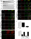
Point mutations in the transmembrane domain of PINK1 partially inhibit its proteolytic cleavage. (a) Amino acids throughout the predicted transmembrane domain of PINK1 were mutated to phenylalanine (amino acids 91–98) or tryptophan (amino acids 99–110). (b) HeLa cells transfected with the indicated PINK1-YFP mutants were treated with either DMSO, 10 µM CCCP, or 10 µM MG132 for 3 h. 20 µg cell lysates were subjected to SDS-PAGE and immunoblotting using antibodies against PINK1 and tubulin. (c) The band intensity of FL PINK1 in DMSO- or CCCP-treated lanes from b was densitometrically measured using Multi Gauge (Fujifilm). After corrections for background and loading, the band intensity ratio of DMSO/CCCP-treated samples for each PINK1 mutant was measured. (d) YFP-tagged WT PINK1 and PINK1 R98F mutants were transfected into HeLa cells. Cells were stained with MitoTracker red before treatment with 10 µM CCCP for 3 h and analyzed by confocal microscopy. (bottom) Enlarged views of the boxed areas are shown. Bars, 20 µm. Green arrowheads, FL and ΔMTS-PINK1; red arrowheads, 52-kD PINK1.
Figure 4.
Figure 4.
The PINK1 R98F mutant resistant to PARL-mediated cleavage is located inside mitochondria. (a) Mitochondria isolated from HeLa cells transfected with YFP-tagged PINK1 R98F were incubated with various concentrations of PK for 30 min on ice and immunoblotted for PINK1, Tom20, Cyt c, AIF, and Hsp70. Green arrowhead, FL and ΔMTS-PINK1; red arrowhead, 52-kD PINK1. (b) HeLa cells transfected with mito-YFP were treated with DMSO or 10 µM CCCP for 3 h followed by incubation in either PBS alone or PBS containing 0.005% digitonin or 0.25% Triton X-100 (TX-100). Cells were immunostained using antibodies against Tom20 and Cyt c and analyzed by confocal microscopy. (c) HeLa cells were transfected with YFP-tagged WT PINK1 or PINK1 R98F mutant for 18 h. Cells were then treated with DMSO or CCCP for 3 h, permeabilized, and immunostained with the indicated antibodies. Images were taken by confocal microscopy. (d) HeLa cells (≥150/condition) stained in c were counted for GFP immunofluorescence. Counting results were represented as mean ± SEM from four replicates. (e) HeLa cells cotransfected with YFP-tagged PINK1 R98F mutant and mCherry-Parkin were incubated with either DMSO or 10 µM CCCP for 1 h followed by confocal imaging. (f) PINK1 KO MEFs transfected with YFP-tagged WT PINK1 or PINK1 R98F mutant together with mCherry-Pax were treated with DMSO or 10 µM CCCP for 3 h, and cells were counted for mitochondrial translocation of Parkin (≥50 cell counts for each sample). Counting results were represented as mean ± SEM from four replicates. Bars, 20 µm.
Figure 5.
Figure 5.
Model for PINK1 import and processing. OM, outer membrane; IM, inner membrane.

References

    1. Cipolat S., Rudka T., Hartmann D., Costa V., Serneels L., Craessaerts K., Metzger K., Frezza C., Annaert W., D’Adamio L., et al. 2006. Mitochondrial rhomboid PARL regulates cytochrome c release during apoptosis via OPA1-dependent cristae remodeling. Cell. 126:163–175 10.1016/j.cell.2006.06.021 - DOI - PubMed
    1. Clark I.E., Dodson M.W., Jiang C., Cao J.H., Huh J.R., Seol J.H., Yoo S.J., Hay B.A., Guo M. 2006. Drosophila pink1 is required for mitochondrial function and interacts genetically with parkin. Nature. 441:1162–1166 10.1038/nature04779 - DOI - PubMed
    1. Gautier C.A., Kitada T., Shen J. 2008. Loss of PINK1 causes mitochondrial functional defects and increased sensitivity to oxidative stress. Proc. Natl. Acad. Sci. USA. 105:11364–11369 10.1073/pnas.0802076105 - DOI - PMC - PubMed
    1. Geisler S., Holmström K.M., Skujat D., Fiesel F.C., Rothfuss O.C., Kahle P.J., Springer W. 2010. PINK1/Parkin-mediated mitophagy is dependent on VDAC1 and p62/SQSTM1. Nat. Cell Biol. 12:119–131 10.1038/ncb2012 - DOI - PubMed
    1. Greene J.C., Whitworth A.J., Kuo I., Andrews L.A., Feany M.B., Pallanck L.J. 2003. Mitochondrial pathology and apoptotic muscle degeneration in Drosophila parkin mutants. Proc. Natl. Acad. Sci. USA. 100:4078–4083 10.1073/pnas.0737556100 - DOI - PMC - PubMed

Publication types