. 2010 Dec 14;107(50):21848-53.
doi: 10.1073/pnas.1011756107. Epub 2010 Nov 29.

Monosynaptic circuit tracing in vivo through Cre-dependent targeting and complementation of modified rabies virus

Affiliations

Monosynaptic circuit tracing in vivo through Cre-dependent targeting and complementation of modified rabies virus

Nicholas R Wall et al. Proc Natl Acad Sci U S A. .

Abstract

We describe a powerful system for revealing the direct monosynaptic inputs to specific cell types in Cre-expressing transgenic mice through the use of Cre-dependent helper virus and a modified rabies virus. We generated helper viruses that target gene expression to Cre-expressing cells, allowing us to control initial rabies virus infection and subsequent monosynaptic retrograde spread. Investigators can use this system to elucidate the connections onto a desired cell type in a high-throughput manner, limited only by the availability of Cre mouse lines. This method allows for identification of circuits that would be extremely tedious or impossible to study with other methods and can be used to build subcircuit maps of inputs onto many different types of cells within the same brain region. Furthermore, by expressing various transgenes from the rabies genome, this system also has the potential to allow manipulation of targeted neuronal circuits without perturbing neighboring cells.


Conflict of interest statement

The authors declare no conflict of interest.

Figures
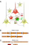
Fig. 1.
Fig. 1.
Using a Cre-dependent helper virus to target rabies virus infection and monosynaptic retrograde spread. (A) Cre-dependent helper virus (green) is injected into the brain of a mouse that expresses Cre in a specific cell type (Cre+). Following Cre recombination, this helper virus expresses two proteins that are necessary for subsequent rabies virus infection (TVA) and monosynaptic retrograde spread (the rabies glycoprotein B19G). TVA is an avian receptor protein that confers infection capability to rabies virus pseudotyped with the avian sarcoma leucosis virus glycoprotein EnvA (red). EnvA-pseudotyped rabies virus is incapable of infecting mammalian neurons in the absence of TVA, restricting rabies virus infection to TVA-expressing cells. The rabies virus has its own glycoprotein gene deleted from its genome, which renders the virus incapable of spreading retrogradely in the absence of another source of rabies glycoprotein. The helper virus complements this deficiency, allowing the rabies virus to spread one synapse retrogradely. This presynaptic neuron population does not express the rabies glycoprotein, so the rabies virus cannot spread any further, limiting rabies virus infection to a small population of Cre-expressing cells and their directly presynaptic partners. (B) Cre-dependent, AAV serotype 9 helper virus vectors were generated to express either GFP and TVA (AAV9-pEF1α-FLEX-GT; Upper) or TVA and Rabies G (AAV9-pEF1α-FLEX-GTB; Lower) in targeted cell types. Cre-dependence was conferred using the FLEX double-floxed system, in which Cre recombinase acts on mutually exclusive sets of lox sites [lox2272 (purple triangles) and loxP (red triangles)] to lock the reverse complemented gene sequence into the proper orientation for transcription. Start ATGs were removed from all genes, and a single Kozak sequence was added outside of the double-floxed cassette, to eliminate any remaining leak expression. To express multiple genes under the control of a single promoter, coding sequences were linked by foot-and-mouth disease virus type 2A elements (blue boxes). (C) The rabies virus genome has been modified such that the glycoprotein gene (G) is replaced with a fluorescent reporter, mCherry. The rabies virus is then pseudotyped with EnvA to produce (EnvA)SAD-ΔG-mCherry.
Fig. 2.
Fig. 2.
Cre-dependent recombination of helper virus gene cassette. The helper viruses carry reverse-complemented gene sequences flanked by two mutually exclusive sets of lox sites, termed FLEX (13). Cre can act on either pair of lox sites, lox2272 (purple triangles) or loxP (red triangles), but cannot mediate recombination across different types of lox sites. Through Cre-mediated inversion of antiparallel lox sites, followed by Cre-mediated deletion of identical parallel lox sites, this double-floxed system stabilizes the Cre-dependent cassette in the proper orientation, allowing for high levels of gene expression.
Fig. 3.
Fig. 3.
Helper virus mediation of rabies virus infection and spread in the cerebellum of L7-Cre mice. (A–C) A helper virus expressing Cre-dependent eGFP and TVA (AAV9-pEF1α-FLEX-GT) was injected into the cerebellum of L7-Cre mice (A and B) and WT mice (C) and allowed to express for 3 wk, either in the absence of rabies virus (A) or followed by injection of rabies virus expressing mCherry [(EnvA)SAD-ΔG-mCherry] (B and C). (A) As expected, the Cre-dependent AAV expresses GFP almost exclusively in Purkinje cells in the L7-Cre mouse. (B) (EnvA)SAD-ΔG-mCherry can only infect cells that are expressing GFP and TVA. In the absence of rabies glycoprotein, the rabies virus is unable to spread from the Purkinje cells to the presynaptic granule cells, labeled densely by blue DAPI stain directly below the Purkinje cell layer. The top row of green Purkinje cells is outside the core region of the rabies virus injection, whereas the bottom row is within the boundary of the rabies virus injection sphere. (C) Cre-dependent AAV does not detectably express GFP, and (EnvA)SAD-ΔG-mCherry is incapable of infecting neurons in the WT animal, indicating that the Cre-dependent AAV effectively restricts TVA expression to only Cre-positive cells. (D–F) A Cre-dependent AAV expressing TVA and rabies glycoprotein (AAV9-pEF1α-FLEX-GTB) (eGFP is in the genome but not detectably expressed) was injected into the cerebellum of L7-Cre mice (D and E) and WT mice (F), followed 3 wk later by injection of (EnvA)SAD-ΔG-mCherry. (D) (EnvA)SAD-ΔG-mCherry can directly infect Purkinje cells and spread transsynaptically to directly presynaptic interneurons and granule cells in the presence of TVA and rabies G in the L7-Cre mouse. The rabies label in D–F is visualized using the Redhot lookup table in F and overlaid onto the DAPI signal (blue), which is used to visualize cerebellar layers. (E) (EnvA)SAD-ΔG-mCherry can spread from directly infected Purkinje cells across the climbing fiber synapse, and can directly label presynaptic cells in the inferior olive in the L7-Cre mouse. (F) As expected, no rabies virus infection is detectable in the WT animal, confirming that TVA expression is properly restricted in the absence of Cre. (Scale bars: 100 μm.)
Fig. 4.
Fig. 4.
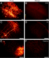
Rabies virus labels direct inputs to PV+ cells in the barrel cortex of PV-Cre mice. Helper virus (AAV9-pEF1α-FLEX-GTB) and rabies virus [(EnvA)SAD-ΔG-mCherry] were injected into the barrel cortex of PV-Cre mice or into WT control animals. mCherry signal is visualized using the Redhot lookup table in B. (A) Rabies virus can infect PV cells and their retrogradely connected partners in the primary somatosensory cortex. (B) In contrast, rabies infection is almost completely absent in the WT animal. (C and D) Retrogradely infected cells can be detected in secondary somatosensory cortex in the PV-Cre mouse (C), but never in the WT animal (D). (E) Retrograde label in the thalamus of PV-Cre mice. Both somata and dense axon termini are reliably detected in thalamic nuclei VPm and Po, which are known to project into barrel fields of the primary somatosensory cortex. Axon terminal labeling arises from corticothalamic projection cells that are retrogradely labeled in A. (F) As expected, there is no mCherry signal in the thalamus of WT animals. (Scale bars: 100 μm.)

References

    1. Luo L, Callaway EM, Svoboda K. Genetic dissection of neural circuits. Neuron. 2008;57:634–660. - PMC - PubMed
    1. Petreanu L, Huber D, Sobczyk A, Svoboda K. Channelrhodopsin-2-assisted circuit mapping of long-range callosal projections. Nat Neurosci. 2007;10:663–668. - PubMed
    1. Braz JM, Rico B, Basbaum AI. Transneuronal tracing of diverse CNS circuits by Cre-mediated induction of wheat germ agglutinin in transgenic mice. Proc Natl Acad Sci USA. 2002;99:15148–15153. - PMC - PubMed
    1. Maskos U, Kissa K, St Cloment C, Brûlet P. Retrograde trans-synaptic transfer of green fluorescent protein allows the genetic mapping of neuronal circuits in transgenic mice. Proc Natl Acad Sci USA. 2002;99:10120–10125. - PMC - PubMed
    1. DeFalco J, et al. Virus-assisted mapping of neural inputs to a feeding center in the hypothalamus. Science. 2001;291:2608–2613. - PubMed

Publication types

LinkOut - more resources