

Case Reports
. 2011 Jul;20 Suppl 2(Suppl 2):S248-52.
doi: 10.1007/s00586-010-1645-x.
Epub 2010 Dec 1.
Complex 360°-reconstruction and stabilization of the cervical spine due to osteomyelitis
Affiliations
- PMID: 21116660
- PMCID: PMC3111505
- DOI: 10.1007/s00586-010-1645-x
Item in Clipboard
Case Reports
Complex 360°-reconstruction and stabilization of the cervical spine due to osteomyelitis
Eur Spine J.
2011 Jul.
Abstract
Osteomyelitis of the cervical spine may lead to profound bony destruction. The presented case developed multilevel osteomyelitic destruction of the cervical spine after decompression due to cervical myelopathy. He could be cured by a multiple-stage procedure: step one: debridement and removal of all anterior implants with vacuum-assisted closure combined with dorsal instrumentation from C0 to T3; step two: anterior reconstruction with expandable titanium cages and plate. The patient regained walking with the aid of a walking frame. The following recommendations are given: multiple stage procedure, extensive debridement and stabilization via an anterior and posterior approach, use of titanium implants.
Figures

July 2008: initial MRI depicting marked spinal stenosis C3–C6

August 2008: situation after the primary surgical intervention with anterior cage-fusion and plating C3–C6

October 2008: MRI after five local revisions depicting severe osteomyelitis additionally involving the C6/C7 segment and epidural abscess at the C2 level
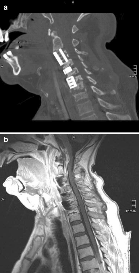
December 2008: a CT showing the situation on admission at our hospital: screws, plates and cages are removed, titanium cages bridging the gaps after corpectomy C3 and partial corpectomy C6 as well as in the disc space C4/C5 are implanted. Note the involvement of the non-fused C7/D1 segment! b Corresponding MRI: note the epidural abscess at the C1–C2 level!
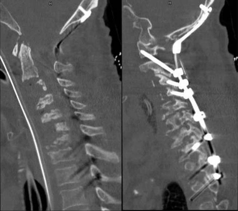
December 2008: a CT after removal of the anteriorly implanted cages and b posterior C0–D3 stabilization
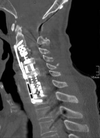
April 2009: CT depicting final anterior stabilization by three expandable titanium cages bridging the gap C2–C4 and C5–D1 and in the disc space C4/C5 and Caspar plating C2–D1

December 2009: 1-year control depicting stable reconstruction of the cervical spine
References
-
- Barnes B, Alexander J, Branch C., Jr Cervical osteomyelitis: a brief review. Neurosurg Focus. 2004;17:E11. - PubMed
Publication types
MeSH terms
LinkOut - more resources
Full Text Sources
Medical
