Review
. 2011 Jul;28(7):1480-99.
doi: 10.1007/s11095-010-0325-1. Epub 2010 Nov 30.

Protein microarrays: novel developments and applications

Affiliations
Review

Protein microarrays: novel developments and applications

Luis Berrade et al. Pharm Res. 2011 Jul.

Abstract

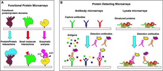
Protein microarray technology possesses some of the greatest potential for providing direct information on protein function and potential drug targets. For example, functional protein microarrays are ideal tools suited for the mapping of biological pathways. They can be used to study most major types of interactions and enzymatic activities that take place in biochemical pathways and have been used for the analysis of simultaneous multiple biomolecular interactions involving protein-protein, protein-lipid, protein-DNA and protein-small molecule interactions. Because of this unique ability to analyze many kinds of molecular interactions en masse, the requirement of very small sample amount and the potential to be miniaturized and automated, protein microarrays are extremely well suited for protein profiling, drug discovery, drug target identification and clinical prognosis and diagnosis. The aim of this review is to summarize the most recent developments in the production, applications and analysis of protein microarrays.


Figures

Fig. 1
Fig. 1
Common formats used for the preparation of protein microarrays. Functional protein microarrays (A) are used to study and identify new molecular interactions between proteins, small molecules or enzyme substrates, for example. Protein detecting microarrays (B) are used to identify proteins from complex mixtures. In the sandwich format (B, left), captured proteins are detected by a secondary antibody typically labeled with a fluorescent dye to facilitate detection and quantification. In contrast to antibody microarrays, lysate microarrays (B, right) are typically immobilized onto nitrocellulose-coated glass slides (FAST slides) and detected using fluorescent-labeled solution-phase specific antibodies.
Fig. 2
Fig. 2
Quantitative interaction networks of tyrosine kinases associated with the Erb family of receptors, which was determined using protein microarrays displaying 96 SH2 and 37 PTB domains. The SH2 and PTB protein domains were probed with fluorescently labeled phosphopeptides representing the different tyrosine phosphoryaltion sites on the Erb kinases. The readout of peptide binding was monitored and quantified by fluorescence. The interaction maps (bottom panel) were constructed from the quantitative interaction data (156). Reprinted from reference (156) with permission from Elsevier.
Fig. 3
Fig. 3
Site-specific and covalent immobilization of a functional protein onto a chemically modified surface using a chemoselective ligation reaction.
Fig. 4
Fig. 4
Principle for site-specific protein immobilization using an active site-directed capture ligand approach.
Fig. 5
Fig. 5
A Site-specific immobilization of cutinase-fusion proteins using an active site-directed capture ligand. B Structure of F. solani cutinase enzyme free and bound to the inhibitor n-undecyl-O-methyl phosphonate chloride. The inhibitor is covalently bound through the side-chain hydroxyl group of the Ser120 residue, which is located at the active site of the enzyme (113).
Fig. 6
Fig. 6
Site-specific immobilization of proteins onto solid supports through protein trans-splicing (85). Maltose binding protein (MPB) was directly immobilized from (a) soluble cellular fraction of E. coli cells over-expressing MBP-IN, and (b) MBP-IN expressed in vitro using an in vitro trascription/traslation expression system. MBP was detected using a fluorescent-labeled specific antibody.
Fig. 7
Fig. 7
In situ methods for protein arraying by PISA (A), NAPPA (B) and puromycin-capture from RNA arrays (C).

References

    1. Tomizaki KY, Usui K, Mihara H. Protein-detecting microarrays: current accomplishments and requirements. Chembiochem. 2005;6(5):782–799. - PubMed
    1. Wolf-Yadlin A, Sevecka M, MacBeath G. Dissecting protein function and signaling using protein microarrays. Curr Opin Chem Biol. 2009;13(4):398–405. - PMC - PubMed
    1. Weinrich D, Jonkheijm P, Niemeyer CM, Waldmann H. Applications of protein biochips in biomedical and biotechnological research. Angew Chem Int Ed Engl. 2009;48(42):7744–7751. - PMC - PubMed
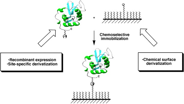
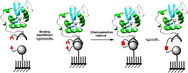
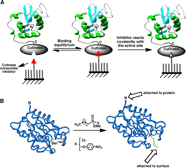
    1. Camarero JA. Recent developments in the site-specific immobilization of proteins onto solid supports. Biopolymers. 2008;90(3):450–458. - PubMed
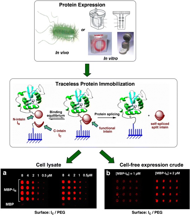
    1. Coleman MA, Hoeprich P, Beernink P, Camarero JA. Cell-free protein expression screening and protein immobilization using protein microarrays. In: Kudlicki W, Katzen F, Bennett R, editors. Cell Free Expression Systems Landes Bioscience Publishers; 2007.

Publication types

Substances