. 2011:699:67-84.
doi: 10.1007/978-1-61737-950-5_4.

Quantum dots for quantitative flow cytometry

Affiliations

Quantum dots for quantitative flow cytometry

Tione Buranda et al. Methods Mol Biol. 2011.

Abstract

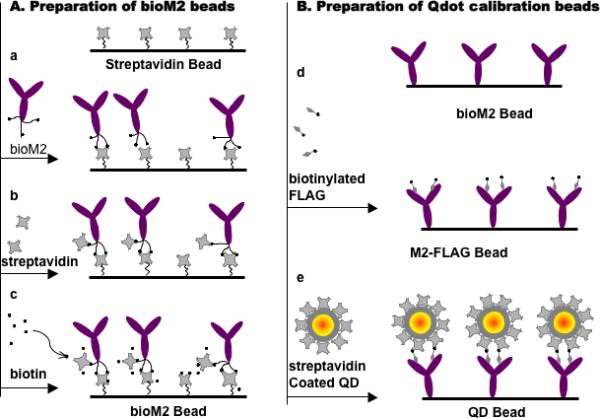
In flow cytometry, the quantitation of fluorophore-tagged ligands and receptors on cells or at particulate surfaces is achieved by the use of standard beads of known calibration. To the best of our knowledge, only those calibration beads based on fluorescein, EGFP, phycoerythyrin and allophycocyanine are readily available from commercial sources. Because fluorophore-based standards are specific to the selected fluorophore tag, their applicability is limited to the spectral region of resonance. Since quantum dots can be photo-excited over a continuous and broad spectral range governed by their size, it is possible to match the spectral range and width (absorbance and emission) of a wide range of fluorophores with appropriate quantum dots. Accordingly, quantitation of site coverage of the target fluorophores can be readily achieved using quantum dots whose emission spectra overlaps with the target fluorophore.This chapter focuses on the relevant spectroscopic concepts and molecular assembly of quantum dot fluorescence calibration beads. We first examine the measurement and applicability of spectroscopic parameters, ε, φ, and %T to fluorescence calibration standards, where ε is the absorption coefficient of the fluorophore, φ is the quantum yield of the fluorophore, and %T is the percent fraction of emitted light that is transmitted by the bandpass filter at the detector PMT. The modular construction of beads decorated with discrete quantities of quantum dots with defined spectroscopic parameters is presented in the context of a generalizable approach to calibrated measurements of fluorescence in flow cytometry.


Figures

Fig. 1
Fig. 1
Summary of the molecular assembly of quantum dot fluorescence calibration beads on streptavidin coated beads. (a) Addition of biotinylated M2 antiFLAG antibodies (bioM2) to 6.7 μm beads. (b) Elimination of free biotin sites with soluble streptavidin prevents direct binding by of qdots. (c) Biotin is used to block streptavidin-biotinylated FLAG peptide interactions. (d) Addition and capture of biotinylated FLAG peptides by bioM2 antibodies. (e) Addition and capture of streptavidin qdots with biotinylated FLAG peptides tethered to bioM2.
Fig. 2
Fig. 2
Comparison of absorbance (A), and uncorrected excitation spectra (B) of quantum dots and fluorescein: (a) Fluorescein, (b) QD525 lot# 1005-0045, and (c) QD585 lot# 0905-0031. The samples’ absorbances were matched at 488 nm by design. (Fig. 2A is reprinted from Analytical Biochemistry , Vol 364, Wu, Y., Lopez, G. P., Sklar, L. A. and Buranda, T. Spectroscopic characterization of streptavidin functionalized quantum dots. 193-203, (2007) with permission from Elsevier).
Fig. 3
Fig. 3
Correcting excitation spectra for the lamp's spectral distribution (Lλ). (A) Overlay of fluorescein's uncorrected excitation spectra with the absorption spectrum. (B) Intensity versus wavelength plots of: (a) the spectral distribution of the Xenon arc lamp used for sample excitation. The lamp profile was derived from (A), by dividing the Lλ-uncorrected emission spectrum of fluorescein by its absorption spectrum; (b) Lλ-uncorrected excitation spectrum of QD525; (c) Lλ-QD525 excitation corrected spectrum. (C) Overlay of Lλ-corrected spectra of (a) fluorescein, (b) QD525, and (c) QD585. (Fig. 3C is reprinted from Analytical Biochemistry , Vol 364, Wu, Y., Lopez, G. P., Sklar, L. A. and Buranda, T. Spectroscopic characterization of streptavidin functionalized quantum dots. 193-203, (2007) with permission from Elsevier).
Fig. 4
Fig. 4
Plot of quantum yields of (a) fluorescein, (b) QD525 lot# 1005-0045, (c) QD585 lot# 0905-0031, and (d) QD605 versus excitation wavelength. The quantum yields were calculated as described Equation. 3. The lines are data derived from excitation spectra while data points represent data derived from integrated intensity. (Reprinted from Analytical Biochemistry , Vol 364, Wu, Y., Lopez, G. P., Sklar, L. A. and Buranda, T. Spectroscopic characterization of streptavidin functionalized quantum dots. 193-203, (2007) with permission from Elsevier).
Fig. 5
Fig. 5
Normalized emission spectra of fluorescein, rhodamine B and quantum dots. Intensity maxima of, QD525 lot# 1005-0045 and QD585 lot# 0905-0031 correspond to their quantum yields relative to fluorescein and rhodamine B (33). Dark and light (green and orange in the electronic version) bars represent BP filters used in a standard flow cytometer (530/30 BP for the FL1 channel, and 585/42 BP for the FL2 channel). The resonance overlap between the BP filters and emission spectra regulates the amount of light that is transmitted or rejected by the BP filter: 28% of fluorescein emission is transmitted to the FL1 channel compared to 38% of QD525, and 65% of QD585 emission is transmitted to the FL2 channel. (Reprinted from Analytical Biochemistry , Vol 364, Wu, Y., Lopez, G. P., Sklar, L. A. and Buranda, T. Spectroscopic characterization of streptavidin functionalized quantum dots. 193-203, (2007) with permission from Elsevier).
Fig. 6
Fig. 6
(A) Flow cytometry histograms of calibration beads bearing a wide range of site densities of QD585 as a result of mixing 1 million bioM2beads with 0, 1 nM, 3 nM, 10 nM, and 30 nM QD585 dots (see Subheading 3.3.). (B) Linear plot of Qdot sites on calibration beads versus the corresponding mean channel fluorescence (MCF) reading. (Reprinted from Analytical Biochemistry , Vol 364, Wu, Y., Campos, S. K., Lopez, G. P., Ozbun, M. A., Sklar, L. A., and Buranda, T. The development of quantum dot calibration beads and quantitative multicolor bioassays in flow cytometry and microscopy. 180-192, (2007) with permission from Elsevier).
Fig. 7
Fig. 7
Simultaneous two-color measurement of eGFP/Qdot 585 nm (QD585) double-stained Human papillomavirus (HPV) particles on A431 cells using flow cytometry and microscopy. (A) Flow cytometry measurement of GFP fluorescence. Solid histogram (MCF ≈ 100) corresponds to cells bearing HPV particles while open histogram (MCF ≈ 50) represents negative control cells with no virus. (B) Fluorescence measurement of QD585. Solid histogram (MCF ≈ 1100) represents cells bearing HPV particles stained with QD585, and open histogram (MCF ≈ 100) represents negative control cells (no virus). (C) Confocal images of pseudovirion (PsV)-bearing A431 cells prepared under similar conditions to flow cytometry measurements as in (A). (D) Confocal images of the same cells after 5 scans show the near complete phototobleaching of eGFP. (E) TEM micrograph of QD585 dots on transparent biotinylated PsV particles, delineated by a random honeycomb grid scaled to fit the size of 60 nm PsV. Insert shows PsV negatively stained with uranyl acetate. The bar scale is 100 nm for both micrographs. (F) Frequency distribution histogram of Qdots/PsV-mean: 11±5. (Fig. 7 A-D is reprinted from Analytical Biochemistry , Vol 364, Wu, Y., Campos, S. K., Lopez, G. P., Ozbun, M. A., Sklar, L. A., and Buranda, T. The development of quantum dot calibration beads and quantitative multicolor bioassays in flow cytometry and microscopy. 180-192, (2007) with permission from Elsevier).

References

    1. Sklar LA. Flow Cytometry in Biotechnology. Oxford University Press; New York, NY.: 2005.
    1. Nolan JP, Sklar LA. The emergence of flow cytometry for sensitive, real-time measurements of molecular interactions. Nat Biotechnol. 1998;16:633–8. - PubMed
    1. Wang L, Gaigalas AK, Abbasi F, Marti G, Vogt R, Schwartz A. Quantitating Fluorescence Intensity From Fluorophores: Practical Use of MESF Values. J Res Natl Inst Stand Technol. 2002;107:339–53. - PMC - PubMed
    1. Schwartz A, Wang A, Early E, et al. Quantitating Fluorescence Intensity from Fluorophore: the definition of MESF Assignment. J Res Natl Inst Stand Technol. 2002;107:83–91. - PMC - PubMed
    1. Wu Y, Campos SK, Lopez GP, Ozbun MA, Sklar LA, Buranda T. The development of quantum dot calibration beads and quantitative multicolor bioassays in flow cytometry and microscopy. Anal Biochem. 2007;364:180–92. - PMC - PubMed

Publication types

LinkOut - more resources