. 2011 Feb;79(2):537-46.
doi: 10.1002/prot.22901.

A mechanism by which binding of the broadly neutralizing antibody b12 unfolds the inner domain α1 helix in an engineered HIV-1 gp120

Affiliations

A mechanism by which binding of the broadly neutralizing antibody b12 unfolds the inner domain α1 helix in an engineered HIV-1 gp120

Ali Emileh et al. Proteins. 2011 Feb.

Abstract

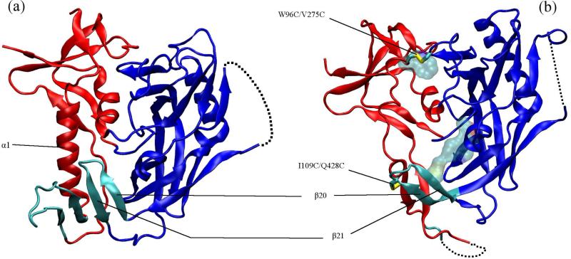
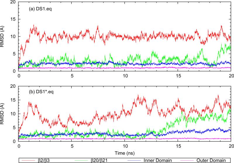
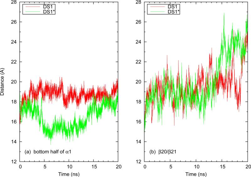
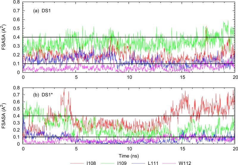
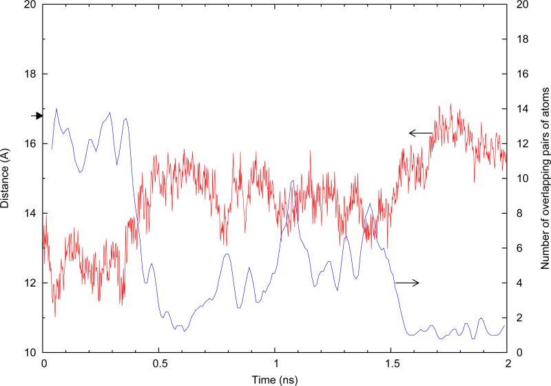
Using all-atom simulations, we examine the role of the I109C/Q428C disulfide "stitch" in altering the conformational distribution of engineered HIV-1 gp120 core relevant for binding of the broadly neutralizing recombinant antibody b12. In particular, we propose that the I109C/Q428C stitch results in a conformational distribution favoring an unfolded inner-domain α1-helix upon binding of b12. Using targeted molecular dynamics, we show that folded α1 in the b12-bound conformation of gp120 is stable both with and without the stitch, but that with folded α1, the stitch requires an orientation of the β20/β21 sheet that is sterically incompatible with b12 binding. Forcing β20/β21 into the orientation displayed by the b12-bound conformation after folding α1 with the stitch intact results in partial unfolding of α1, whereas without the stitch, β20/β21 reorientation does not affect the conformation of α1. These findings collectively support the hypothesis that the disulfide stitch shifts the conformational distribution of α1 to the unfolded state, meaning an unfolded α1 is not a strict requirement of the b12-bound conformational ensemble of gp120's lacking the I109C/Q428C stitch.


Figures

FIG. 1
FIG. 1
(a) CD4-bound (PDB 1GC1) and (b) b12-bound (PDB 2NY7) conformations of gp120. The inner domain is red, outer domain blue and the bridging sheet cyan. In (b) licorice rendering shows the two disulfide bridges (hydrogen atoms not shown) between the inner and outer domain and the residues shown in transparent van der Waals are the other stabilizing mutations (M95W, T257S, S375W, A433M). The dashed lines show the unresolved V4 domain in both structures and also parts of the β2/β3 domain and base of the V1/V2 loop in the b12-bound conformation.
FIG. 2
FIG. 2
Cα RMSD trace of different gp120 domains after alignment using the outer domain: (a) disulfide-“stitched” structure (b) non-stitched structure.
FIG. 3
FIG. 3
Distance of (a) α1 and (b) β20/β21 from the Cα atom of core residue S257 during equilibration of DS1 and DS1*. β20/β21 is represented by the Cα atom of residue G431 at its tip. α1 is represented by the center of mass of residues 108-112.
FIG. 4
FIG. 4
Hydrophobic residues in and around the putative F43 pocket in the DS1* structure. Residues in red make up the F43 pocket (5). Residues shown in yellow are hydrophobic residues within 10 Å of residue 382 and taken to be close to the pocket. The position of the β20/β21 domain at the beginning (red) and end (blue) of the DS1* equilibration is shown in cartoon rendering.
FIG. 5
FIG. 5
Evolution of fractional surface accessible area (FSASA) for residues at the bottom of α1. The dashed lines at 0.1 and 0.4 show the limiting values for definition of buried and exposed residues.
FIG. 6
FIG. 6
Equilibrium backbone RMSD evolution after removal of the TMD forces. Each frame in the trajectory is aligned over the first frame of equilibration (i.e. last frame of TMD forcing) using the outer domain backbone (details explained in the previous section).
FIG. 7
FIG. 7
Movements of the β20/β21 strand relative to the core as measured by the distance G431.CA-T257.CA, during TMD and post-TMD equlibrations. The small arrow at 16.95 Å shows the value of the metric in the CD4-bound crystal structure (1GC1) of gp120. The vertical line indicates the transition from TMD to equilibration.
FIG. 8
FIG. 8
Number of overlapping pairs of atoms resulting from overlaying b12 on α1-folded gp120. The raw data were smoothed using a triangular average method with a sampling window size of 0.5 ns. The vertical line indicates where the TMD forces are lifted and equilibration is started. The small arrow at n=6 overlaps shows the number of overlaps generated when using gp120 extracted the CD4-bound crystal structure (PDB 1GC1).
FIG. 9
FIG. 9
Distance between Cα atoms of G431 and S257 when β20/β21 is repositioned. The small arrow at 16.8 Å indicates this distance in the 2NY7 crystal structure of stitched gp120. Also plotted, is the number of b12/gp120 overlaps (smoothed with a sampling window size of 0.02 ns).
FIG. 10
FIG. 10
(a) Schematic representation of how β20/β21 was moved when it was pushed away from the hydrophobic core in DS1. Representative frames with a sampling rate of 1 frame per 0.5 ns are shown in tube rendering with the starting state in red and the final state in blue (only the β20/β21 region is depicted, the coordinates of the rest of the molecule are from the starting frame). β20/β21 from the crystal structure is shown in blue cartoon rendering. (a) α1 helix conformation during the TMD run with red showing the domain in the beginning and blue in the end of the simulation. At least 5 hydrogen bonds are destroyed.

Similar articles

Cited by

References

    1. Poignard P, Saphire E, Parren P, Burton D. Gp120: Biologic aspects of structural features. Annu Rev Immunol. 2001;19:253–274. - PubMed
    1. Phogat S, Wyatt R. Rational modifications of hiv-1 envelope glycoproteins for immunogen design. Curr Pharm Design. 2007;13:213–227. - PubMed
    1. Wyatt R, Sodroski J. The hiv-1 envelope glycoproteins: Fusogens, antigens, and immunogens. Science. 1998;280:1884–1888. - PubMed
    1. Weiss C, Levy J, White J. Oligomeric organization of gp120 on infectious human-immunodefficiency-virus type-1 particles. J Virol. 1990;64:5674–5677. - PMC - PubMed
    1. Kwong P, Wyatt R, Robinson J, Sweet R, Sodroski J, Hendrickson W. Structure of an hiv gp120 envelope glycoprotein in complex with the cd4 receptor and a neutralizing human antibody. Nature. 1998;393:648–659. - PMC - PubMed

Publication types

MeSH terms

LinkOut - more resources