. 2011 Feb;218(2):185-90.
doi: 10.1111/j.1469-7580.2010.01325.x. Epub 2010 Nov 30.

Anatomical and functional segments of the deltoid muscle

Affiliations

Anatomical and functional segments of the deltoid muscle

Yoshimasa Sakoma et al. J Anat. 2011 Feb.

Abstract

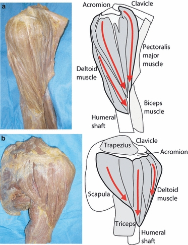
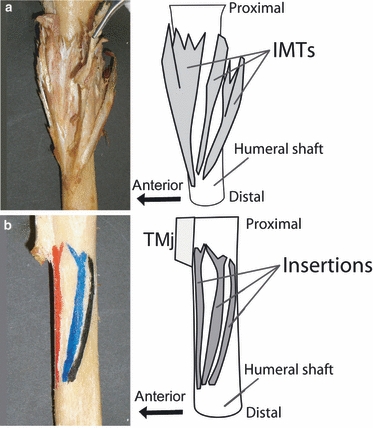
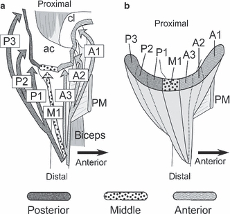
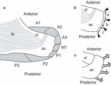
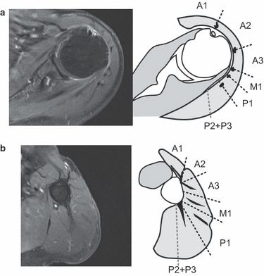
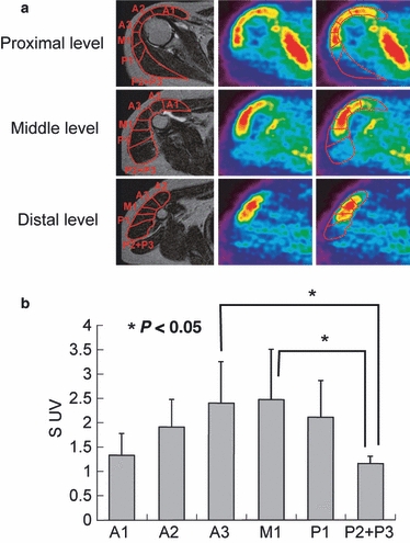
Previous studies showed that the insertion of the intramuscular tendons of the deltoid muscle formed three discrete lines. The purpose of the present study was to establish a new dividing method of the deltoid muscle into various anatomical segments based on the distribution of the intramuscular tendons with their insertions (anatomical study). We further hoped to clarify the relationship between the anatomical segments and their activity pattern assessed by positron emission tomography with [¹⁸F]-2-fluoro-deoxyglucose (FDG-PET; PET study). Sixty cadaveric shoulders were investigated in the anatomical study. Three tendinous insertions of the deltoid muscle to the humerus were identified. Then, the intramuscular tendons were traced from their humeral insertions to the proximal muscular origins. The extent of each anatomical segment of the muscle including its origin and insertion was determined through careful dissection. Six healthy volunteers were examined using FDG-PET for the PET study. PET images were obtained after exercise of elevation in the scapular plane. On the PET images, margins of each anatomical segment of the deltoid muscle were determined using magnetic resonance images. Then, the standardized uptake value in each segment was calculated to quantify its activity. The anatomical study demonstrated that the deltoid muscle was divided into seven segments based on the distribution of its intramuscular tendons. The PET study revealed that the intake of FDG was not uniform in the deltoid muscle. The area with high FDG intake corresponded well to the individual muscular segments separated by the intramuscular tendons. We conclude that the deltoid muscle has seven anatomical segments, which seem to represent the functional units of this muscle.


Figures

Fig. 1
Fig. 1
Surface anatomy of the deltoid muscle. (a) The anterolateral view of the deltoid muscle. The muscle fibers run in the anteroinferior direction (red arrows), which converge the anterior side of the deltoid tubercula. (b) The posterior view of the deltoid muscle.
Fig. 2
Fig. 2
Anatomy of the insertion of the deltoid into the humeral shaft. (a) Three intramuscular tendons insert individually into the humeral shaft (IMTs, intramuscular tendons). (b) The insertions of the intramuscular tendons form three discrete lines (TMj, teres major muscle).
Fig. 3
Fig. 3
(a) Directions of the intramuscular tendons. Arrows indicate the direction of each intramuscular tendon (A1, A2, A3, M1, P1, P2 and P3, intramuscular tendons; ac, acromion; cl, clavicle; PM, pectoralis major muscle). (b) Seven segments at the proximal part of the deltoid muscle on the transverse plane.
Fig. 4
Fig. 4
The landmarks at the origin of the deltoid muscle. (a) Origin of each segment. The border between A1 and A2 locates approximately 5 mm medial from the acromioclavicular joint. There are no particular structures between P2 and P3 at the proximal part of the deltoid (ac, acromion; cl, clavicle; tp, trapezius). (b) There are two bony tubercula at the lateral border of the acromion. These bony tubercula separate the lateral border of the acromion into three facets: a, anterior facet; b, middle facet; c, posterior facet. (c) Four intramuscular tendons originate from the anterior angle, two bony tubercula and posterior angle of the acromion, respectively.
Fig. 5
Fig. 5
Dividing method of the deltoid muscle on MR images. The intramuscular tendons are clearly depicted in a T2-weighted transverse MR image with fat suppression. The deltoid muscle is divided based on the distribution of intramuscular tendons. The straight lines show the border between each muscle segment. The proximal intramuscular tendons locate at the borders of each segment. For the distal insertions, A3, M1 and P1 intramuscular tendons exist at their center. On the other hand, A1, A2 and P3 have their distal intramuscular tendons at the margin of the segments. (a) At proximal deltoid level, (b) At distal deltoid level.
Fig. 6
Fig. 6
(a) Axial MRI and PET images of the proximal, middle and distal levels of the deltoid. At proximal level, the segments divided on MRI well correspond to the dotted FDG intake pattern in the PET images. (b) SUVs of each segment. The standardized uptake values (SUVs) of A3 and M1 are relatively higher than that of other segments. Especially, both A3 and M1 activities were significantly greater than that of P2 + P3 (P< 0.05).

References

    1. Brown JM, Wickham J. Neuromotor coordination of multisegmental muscle during a change in movement direction. J Musculoskelet Res. 2006;10:63–74.
    1. Brown JM, Wickham J, McAndrew DJ, et al. Muscles within muscles: coordination of 19 muscle segments within three shoulder muscles during isometric motor tasks. J Electromyogr Kinesiol. 2007;17:57–73. - PubMed
    1. Cordasco FA, Chen NC, Backus SI, et al. Subacromial injection improves deltoid firing in subjects with large rotator cuff tears. HSS J. 2010;6:30–36. - PMC - PubMed
    1. Fick R. Handbuch der Anatomie und Mekanik der Gelenke. Jena: Gustav Fischer; 1911.
    1. Finni T. Structural and functional features of human muscle-tendon unit. Scand J Med Sci Sports. 2006;16:147–158. - PubMed

Substances

LinkOut - more resources