

Pyrenophora teres: profile of an increasingly damaging barley pathogen
- PMID: 21118345
- PMCID: PMC6640222
- DOI: 10.1111/j.1364-3703.2010.00649.x
Pyrenophora teres: profile of an increasingly damaging barley pathogen
Abstract
Pyrenophora teres, causal agent of net blotch of barley, exists in two forms, designated P. teres f. teres and P. teres f. maculata, which induce net form net blotch (NFNB) and spot form net blotch (SFNB), respectively. Significantly more work has been performed on the net form than on the spot form although recent activity in spot form research has increased because of epidemics of SFNB in barley-producing regions. Genetic studies have demonstrated that NFNB resistance in barley is present in both dominant and recessive forms, and that resistance/susceptibility to both forms can be conferred by major genes, although minor quantitative trait loci have also been identified. Early work on the virulence of the pathogen showed toxin effector production to be important in disease induction by both forms of pathogen. Since then, several laboratories have investigated effectors of virulence and avirulence, and both forms are complex in their interaction with the host. Here, we assemble recent information from the literature that describes both forms of this important pathogen and includes reports describing the host-pathogen interaction with barley. We also include preliminary findings from a genome sequence survey.
Taxonomy: Pyrenophora teres Drechs. Kingdom Fungi; Phylum Ascomycota; Subphylum Pezizomycotina; Class Dothideomycete; Order Pleosporales; Family Pleosporaceae; Genus Pyrenophora, form teres and form maculata.
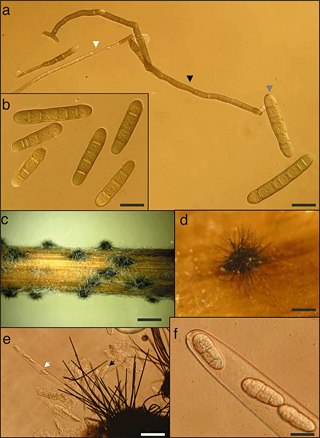
Identification: To date, no clear morphological or life cycle differences between the two forms of P. teres have been identified, and therefore they are described collectively. Towards the end of the growing season, the fungus produces dark, globosely shaped pseudothecia, about 1-2mm in diameter, on barley. Ascospores measuring 18-28µm × 43-61µm are light brown and ellipsoidal and often have three to four transverse septa and one or two longitudinal septa in the median cells. Conidiophores usually arise singly or in groups of two or three and are lightly swollen at the base. Conidia measuring 30-174µm × 15-23µm are smoothly cylindrical and straight, round at both ends, subhyaline to yellowish brown, often with four to six pseudosepta. Morphologically, P. teres f. teres and P. teres f. maculata are indistinguishable.
Host range: Comprehensive work on the host range of P. teres f. teres has been performed; however, little information on the host range of P. teres f. maculata is available. Hordeum vulgare and H. vulgare ssp. spontaneum are considered to be the primary hosts for P. teres. However, natural infection by P. teres has been observed in other wild Hordeum species and related species from the genera Bromus, Avena and Triticum, including H. marinum, H. murinum, H. brachyantherum, H. distichon, H. hystrix, B. diandrus, A. fatua, A. sativa and T. aestivum (Shipton et al., 1973, Rev. Plant Pathol. 52:269-290). In artificial inoculation experiments under field conditions, P. teres f. teres has been shown to infect a wide range of gramineous species in the genera Agropyron, Brachypodium, Elymus, Cynodon, Deschampsia, Hordelymus and Stipa (Brown et al., 1993, Plant Dis. 77:942-947). Additionally, 43 gramineous species were used in a growth chamber study and at least one of the P. teres f. teres isolates used was able to infect 28 of the 43 species tested. However, of these 28 species, 14 exhibited weak type 1 or 2 reactions on the NFNB 1-10 scale (Tekauz, 1985). These reaction types are small pin-point lesions and could possibly be interpreted as nonhost reactions. In addition, the P. teres f. teres host range was investigated under field conditions by artificially inoculating 95 gramineous species with naturally infected barley straw. Pyrenophora teres f. teres was re-isolated from 65 of the species when infected leaves of adult plants were incubated on nutrient agar plates; however, other than Hordeum species, only two of the 65 host species exhibited moderately susceptible or susceptible field reaction types, with most species showing small dark necrotic lesions indicative of a highly resistant response to P. teres f. teres. Although these wild species have the potential to be alternative hosts, the high level of resistance identified for most of the species makes their role as a source of primary inoculum questionable.
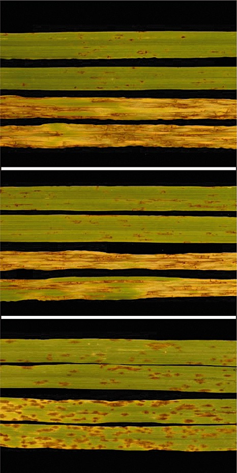
Disease symptoms: Two types of symptom are caused by P. teres. These are net-type lesions caused by P. teres f. teres and spot-type lesions caused by P. teres f. maculata. The net-like symptom, for which the disease was originally named, has characteristic narrow, dark-brown, longitudinal and transverse striations on infected leaves. The spot form symptom consists of dark-brown, circular to elliptical lesions surrounded by a chlorotic or necrotic halo of varying width.
Molecular Plant Pathology © 2010 BSPP and Blackwell Publishing Ltd. No claim to original US Government Works.
Figures
References
-
- Able, A. (2003) Role of reactive oxygen species in the response of barley to necrotrophic pathogens. Protoplasma, 221, 137–143. - PubMed
-
- Abu Qamar, M. , Liu, Z.H. , Faris, J.D. , Chao, S. , Edwards, M.C. , Lai, Z. , Franckowiak, J.D. and Friesen, T.L. (2008) A region of barley chromosome 6H harbors multiple major genes associated with net type net blotch resistance. Theor. Appl. Genet. 117, 1261–1270. - PubMed
-
- Afanasenko, O. , Mironenko, N. , Filatova, O. , Kopahnke, D. , Krämer, I. and Ordon, F. (2007) Genetics of host–pathogen interactions in the Pyrenophora teres f. teres (net form)–barley (Hordeum vulgare) pathosystem. Eur. J. Plant Pathol. 117, 267–280.
-
- Afanasenko, O.S. , Jalli, M. , Pinnschmidt, H.O. , Filatova, O. and Platz, G.J. (2009) Development of an international standard set of barley differential genotypes for Pyrenophora teres f. teres . Plant Pathol. 58, 665–676.
-
- Alcorn, J.L. (1988) The taxonomy of ‘Helminthosporium’ species. Annu. Rev. Phytopathol. 26, 37–56.
Publication types
MeSH terms
Substances
LinkOut - more resources
Full Text Sources
