Comparative Study
. 2010;14(6):R220.
doi: 10.1186/cc9354. Epub 2010 Nov 30.

PD-L1 blockade improves survival in experimental sepsis by inhibiting lymphocyte apoptosis and reversing monocyte dysfunction

Affiliations
Comparative Study

PD-L1 blockade improves survival in experimental sepsis by inhibiting lymphocyte apoptosis and reversing monocyte dysfunction

Yan Zhang et al. Crit Care. 2010.

Abstract

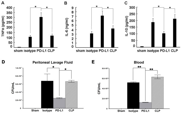
Introduction: Lymphocyte apoptosis and monocyte dysfunction play a pivotal role in sepsis-induced immunosuppression. Programmed death-1 (PD1) and its ligand programmed death ligand-1 (PD-L1) exert inhibitory function by regulating the balance among T cell activation, tolerance, and immunopathology. PD-1 deficiency or blockade has been shown to improve survival in murine sepsis. However, PD-L1 and PD-1 differ in their expression patterns and the role of PD-L1 in sepsis-induced immunosuppression is still unknown.

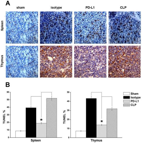
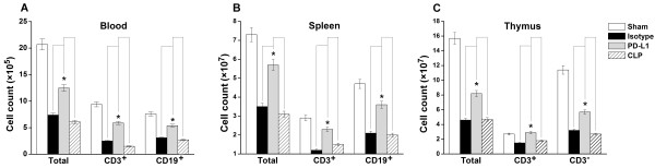
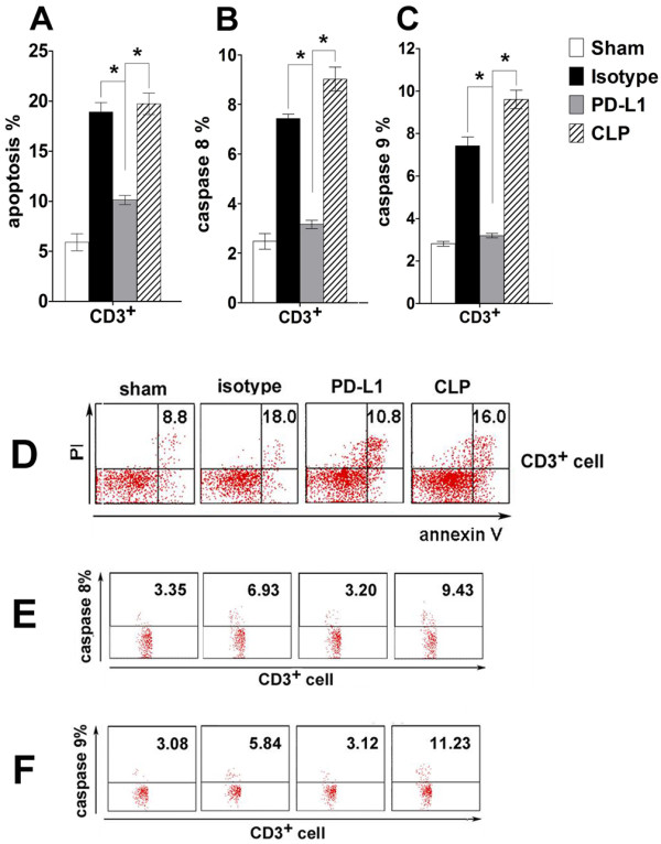
Methods: Sepsis was induced in adult C57BL/6 male mice via cecal ligation and puncture (CLP). The expression of PD-1 and PD-L1 expression on peripheral T cells, B cells and monocytes were measured 24 hours after CLP or sham surgery. Additionally, the effects of anti-PD-L1 antibody on lymphocyte number, apoptosis of spleen and thymus, activities of caspase-8 and caspase-9, cytokine production, bacterial clearance, and survival were determined.

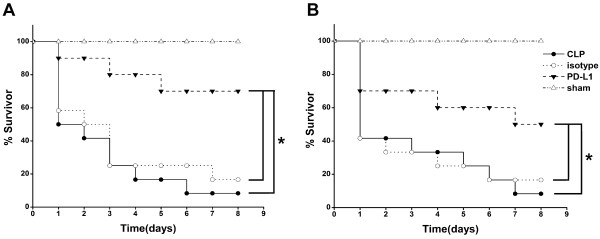
Results: Expression of PD-1 on T cells, B cells and monocytes and PD-L1 on B cells and monocytes were up-regulated in septic animals compared to sham-operated controls. PD-L1 blockade significantly improved survival of CLP mice. Anti-PD-L1 antibody administration prevented sepsis-induced depletion of lymphocytes, increased tumor necrosis factor (TNF)-α and interleukin (IL)-6 production, decreased IL-10 production, and enhanced bacterial clearance.

Conclusions: PD-L1 blockade exerts a protective effect on sepsis at least partly by inhibiting lymphocyte apoptosis and reversing monocyte dysfunction. Anti-PD-L1 antibody administration may be a promising therapeutic strategy for sepsis-induced immunosuppression.


Figures

Figure 1
Figure 1
PD-1 and PD-L1 expression on T cells, B cells and monocytes. (A) Percent of PD-1 expression on CD3+ T cells, CD19+ B cells and CD11b+ monocytes 24 h after CLP (n = 5) or sham control surgery (n = 5). (B) Percent of PD-L1 expression on CD3+ T cells, CD19+B cells and CD11b+ monocytes 24 h after CLP (n = 5) or sham control surgery (n = 5), (C, D) Representative PD-1 and PD-L1 expression on T, B cells and monocytes detected by flow cytometry. * P <0.05, ** P <0.01.
Figure 2
Figure 2
Anti-PD-L1 antibody administration protects mice from sepsis-induced lethality. (A) Anti-PD-L1 antibody pretreatment protected mice from CLP. CLP mice were given 50 μg anti-PD-L1 antibody (n = 18), 50 μg isotype control antibody (n = 12) or 0.2 mL saline intraperitoneally 24 h before CLP surgery. (B) Effect of intraperitoneal anti-PD-L1 antibody treatment given 3 h after CLP. CLP mice were given 50 μg anti-PD-L1 antibody (n = 18), 50 μg isotype control antibody (n = 12) or 0.2 mL saline (n = 12) intraperitoneally 3 h after CLP. Survival was monitored for eight days. Data are shown as the survival percent of animals. *P <0.05.
Figure 3
Figure 3
PD-L1 blockade inhibits cell apoptosis in spleen and thymus. Mice underwent a sham procedure, CLP, CLP plus anti-PD-L1 or isotype administration (n = 5 for each group). Spleen and thymus were harvested 24 h after surgery. (A) Representative sections analyzed by an in situ TUNEL assay. (B) Percent of the TUNEL-positive cells is used to show the cell apoptosis in spleen and thymus of the 4 groups. * P <0.05.
Figure 4
Figure 4
Cell numbers in blood (A), spleen (B) and thymus (C). Mice underwent a sham procedure, CLP, CLP plus anti-PD-L1 administration, or CLP plus isotype administration (n = 5 for each group). Blood, thymus and spleen were harvested 24 h after surgery. The total cell number was counted after lysis of erythrocytes (for spleen and thymus, preparation of a single-cell suspension was required). Lymphocyte numbers (CD3+ T cells, CD3- T cells, CD19+ B cells) were calculated by the total number and percent of lymphocyte subgroups resulting from FACS analysis, respectively. * P <0.05.
Figure 5
Figure 5
Both extrinsic and intrinsic pathways contribute to decreased lymphocyte apoptosis in vivo. Mice underwent sham procedure, CLP, CLP plus anti-PD-L1 antibody administration, or CLP plus isotype administration. Thymus was harvested 24 h after surgery and stained for annexin V and propidium iodide (PI) (A, D) or FITC-labeled IEHD-FMK (B, E) or LEHD-FMK (C, F) which can irreversibly binds to activated caspase-8 or activated caspase-9. (D), (E), and (F) are the representative flow cytometry dot plots. Values in the upper right quadrant indicate the percent of apoptotic cells, caspase-8 or caspase-9 positive cells, respectively. * P <0.05.
Figure 6
Figure 6
Levels of plasma cytokines and bacterial clearance. Mice underwent a sham procedure, CLP, CLP plus anti-PD-L1 administration, or CLP plus isotype control administration (n = 5 for each group). Levels of TNF-α (A), IL-6 (B) and IL-10 (C) were measured 24 h after surgery. Treatment with anti-PDL1 antibody improves bacterial clearance in septic mice. Mice that received anti-PD-L1 antibody exhibited a deceased bacterial burden in peritoneal lavage fluid in comparison with mice that received isotype antibody or saline (D), Mice that received anti-PD-L1 antibody exhibited a deceased bacterial burden in blood in comparison with mice that received isotype antibody or saline (E), * P < 0.05, ** P < 0.01.

References

    1. Deans KJ, Haley M, Natanson C, Eichacker PQ, Minneci PC. Novel therapies for sepsis: a review. J Trauma. 2005;58:867–874. doi: 10.1097/01.TA.0000158244.69179.94. - DOI - PubMed
    1. Riedemann NC, Guo RF, Ward PA. The enigma of sepsis. J Clin Invest. 2003;112:460–467. - PMC - PubMed
    1. Wisnoski N, Chung CS, Chen Y, Huang X, Ayala A. The contribution of CD4+ CD25+ T-regulatory-cells to immune suppression in sepsis. Shock. 2007;27:251–257. doi: 10.1097/01.shk.0000239780.33398.e4. - DOI - PMC - PubMed
    1. Hotchkiss RS, Coopersmith CM, McDunn JE, Ferguson TA. The sepsis seesaw: tilting toward immunosuppression. Nat Med. 2009;15:496–497. doi: 10.1038/nm0509-496. - DOI - PMC - PubMed
    1. Lang JD, Matute-Bello G. Lymphocytes, apoptosis and sepsis: making the jump from mice to humans. Crit Care. 2009;13:109–110. doi: 10.1186/cc7144. - DOI - PMC - PubMed

Publication types