. 2011 Jan;46(1):347-56.
doi: 10.1016/j.mcn.2010.11.003. Epub 2010 Nov 27.

Midbrain dopaminergic axons are guided longitudinally through the diencephalon by Slit/Robo signals

Affiliations

Midbrain dopaminergic axons are guided longitudinally through the diencephalon by Slit/Robo signals

James P Dugan et al. Mol Cell Neurosci. 2011 Jan.

Abstract

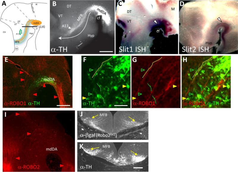
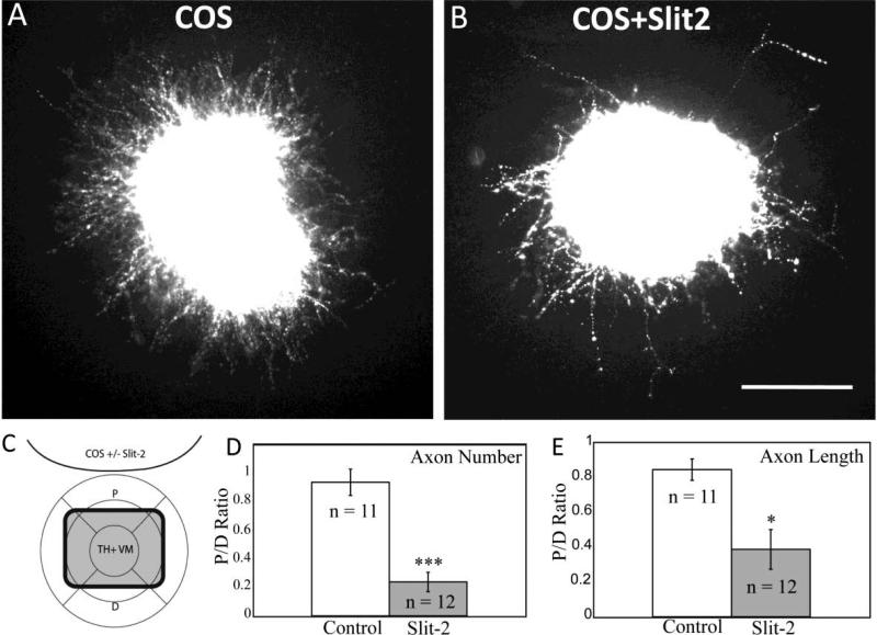
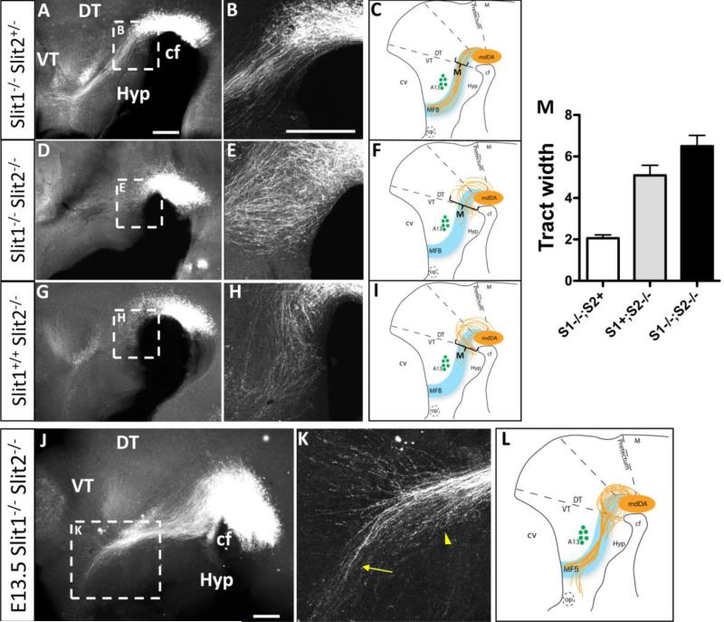
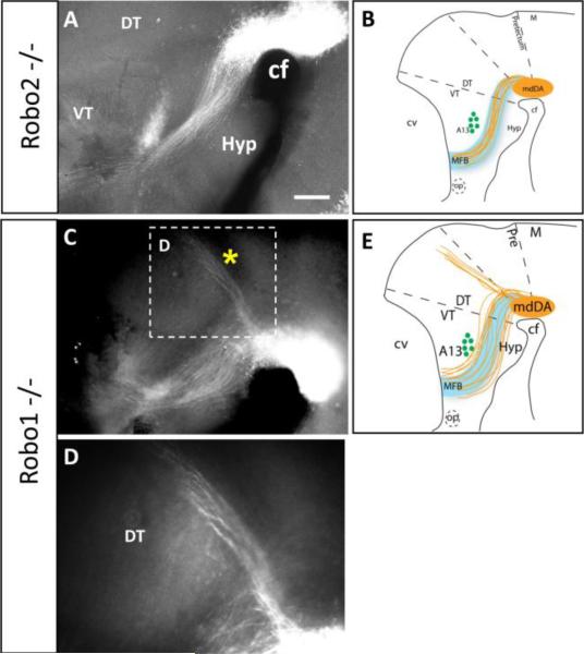
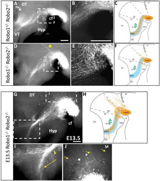
Dopaminergic neurons from the ventral mesencephalon/diencephalon (mesodiencephalon) form vital pathways constituting the majority of the brain's dopamine systems. Mesodiencephalic dopaminergic (mdDA) neurons extend longitudinal projections anteriorly through the diencephalon, ascending toward forebrain targets. The mechanisms by which mdDA axons initially navigate through the diencephalon are poorly understood. Recently the Slit family of secreted axon guidance proteins, and their Robo receptors, have been identified as important guides for descending longitudinal axons. To test the potential roles of Slit/Robo guidance in ascending trajectories, we examined tyrosine hydroxylase-positive (TH+) projections from mdDA neurons in mutant mouse embryos. We found that mdDA axons grow out of and parallel to Slit-positive ventral regions within the diencephalon, and that subsets of the mdDA axons likely express Robo1 and possibly also Robo2. Slit2 was able to directly inhibit TH axon outgrowth in explant co-culture assays. The mdDA axons made significant pathfinding errors in Slit1/2 and Robo1/2 knockout mice, including spreading out in the diencephalon to form a wider tract. The wider tract resulted from a combination of invasion of the ventral midline, consistent with Slit repulsion, but also axons wandering dorsally, away from the ventral midline. Aberrant dorsal trajectories were prominent in Robo1 and Robo1/2 knockout mice, suggesting that an aspect of Robo receptor function is Slit-independent. These results indicate that Slit/Robo signaling is critical during the initial establishment of dopaminergic pathways, with roles in the dorsoventral positioning and precise pathfinding of these ascending longitudinal axons.


Figures

Figure 1
Figure 1
TH+ axons project adjacent to Slit+ tissue, and express Robo1 and 2. A. Schematic representation of the initial mdDA projections through the diencephalon. The nucleus of the mdDA neurons is positioned along the cephalic flexure and extends across the forebrain/midbrain boundary. These TH+ axons project anteriorly in a distinct ventrolateral position through the thalamus. Adjacent to the MFB tract, the A13 cell population serves as a landmark. Black lines marked E-F and G-H indicate section planes in those panels. B. Wild-type whole mount embryo in the CD-1 background immunolabeled with TH. The white arrow represents the normal trajectory of mdDA axons through the thalamus. The tract is labeled MFB. C. Slit1 mRNA expression (purple) is localized around the cephalic flexure, with robust signal at the ventral midline (arrowhead). Fainter graded expression domains are visible in dorsal midbrain, and dorsal and ventral thalamus. D. Slit2 expression is also seen along the ventral midline, overlapping with the expression of Slit1. Very faint expression is also found in the hypothalamus and lateral thalamus. E. Robo1 (red) and TH (green) antibody labeling of a sagittal section of an E12.5 embryo. Robo1 is expressed in an extensive set of longitudinal bundles (E) which transit the mdDA nucleus. F-H. High magnification views of a different section. Some TH cell bodies are Robo1+ (yellow arrowheads), while other TH+ cell bodies do not show detectable Robo1 labeling (green arrowheads). The dotted lines trace a few examples of TH+ axons, of which some are closely associated with Robo1+ bundles (yellow), while other TH+ axons are not (green). I. Robo2 antibody labeling of a sagittal section of an E12.5 embryo. While Robo2 label can be seen in longitudinal fibers in dorsal, anterior, and posterior regions, little to no Robo2 label can be seen in the area of the mdDA or the MFB. J, K. Adjacent tissue sections showing anti-β-galactosidase immunolabeling representing expression from a Robo2lacZ allele (J), in comparison to TH in the MFB (K). Both markers label MFB fibers (yellow arrows). Abbreviations: cf, cephalic flexure; cv, cerebral vesicle; DT, dorsal thalamus; Hyp, hypothalamus; M, midbrain; md, mesodiencephalon; MFB, medial forebrain bundle; op, optic stalk; VT, ventral thalamus. Scale Bars: B and E, 400 μm; (applies to C, D, I); F, 50 μm; (applies to G, H); K, 100 μm (applies to J).
Figure 2
Figure 2
TH+ axons are inhibited by secreted Slit2 in vitro. Ventral midbrain tissue from E12.5 mouse embryos was cultured for two days in collagen gel with aggregates of COS cells, followed by fixation and labeling with TH antibodies. Images are oriented with COS aggregates at the top. A. Control explant with mock-transfected COS cell aggregate. B. Explant challenged with Slit2-expressing COS cell aggregate. C. Schematic depicting TH+ ventral mesencephalic (VM) explant and COS cell aggregate (with or without Slit2 expression). Crosshairs define proximal (P) and distal (D) quadrants with relation to aggregate. Number and length of axons were determined in each quadrant and the ratio P/D was determined for every explant. D. P/D ratio of the number of axons in control explants versus explants cocultured with Slit2 expressing COS cells. E. P/D ratio of the length of axons in control explants versus explants cocultured with Slit2 expressing COS cells. The P/D ratio with Slit2 was significantly decreased compared to control explants, by t-test. ***p<.0001, *p<.05. Scale bar: 100 μm.
Figure 3
Figure 3
Slits organize mdDA projections through the diencephalon, with a dominant role for Slit2. Whole-mount TH immunolabeling and schematics of E12.5 (A-I) and E13.5 (J-M) embryos at low (A,D,G,J) and high (B,E,H,K) magnification. A-C. TH+ axons in Slit1-/-;Slit2+/- double mutants, with projections identical to wildtype controls (compare to Fig 1B). Note that TH+ axons project in an organized and relatively narrow ascending trajectory through the diencephalon toward the telencephalon. Axons avoid the ventral hypothalamus, and project ventral to the A13 landmark. D-F. Slit1-/-; Slit2-/- double homozygotes form a wider tract, with many axons growing within ventral midline tissue near the cephalic flexure (E). In addition, axons have shorter projections with few in the proper direction, and some wandering dorsally. G-I. Slit1+/+; Slit2-/- single mutant embryos are similar to Slit1-/-; Slit2-/- double mutants, with projections looping into the ventral midline (*), extending over a wider dorsoventral breadth, and of shorter length than in control embryos. J-L. By E13.5 the ventral looping into the midline is still prevalent and mdDA neurons continue to project over a wider dorsoventral breadth, invading ventral midline tissue. K. Within the anterior diencephalon, mdDA axons deviate from the tract to project toward the optic stalk (arrow). Axons in the tract converge abnormally (arrowhead). M. Quantitation of the width of the mdDA tract, measured at the point indicated in the schematic summary of each mutant phenotype. Tract width is indicated by arbitrary units consistent between the genotype pools, +/- SEM. Slit1-/-; Slit2+ is a pool of control embryos (n=8) derived from the breeding stock of Slit1 homozygotes, with or without a Slit2- allele. Slit1+; Slit2-/- are a similar pool of embryos with at least one Slit1+ allele (n=6), compared to Slit1-/-; Slit2-/- double mutants (n=7). By ANOVA, the three pools were significantly different. cf, cephalic flexure; DT, dorsal thalamus; Hyp, hypothalamus; M, midbrain; VT, ventral thalamus. Scale bars, in A and B represent 400 μm for low and high magnification, respectively.
Figure 4
Figure 4
Robo single mutants suggest a dominant role for Robo1 in mdDA axon guidance. Whole-mount TH immunolabeling of E13.5 embryos shows Robo2-/- mutant (A) and Robo1-/- mutant phenotypes at low (A,C) and high magnification (D). A. Robo2-/- mutant embryos have normal axonal projections forming a narrow tract through the diencephalon (compare to Fig. 1B). C-E. Robo1-/- mutants are characterized by a dorsoventral broadening of the tract. Most axons continue along the MFB projecting into the telencephalon. However, a pronounced bundle of axons projects from the mdDA nucleus dorsally along the dorsal thalamus-pretectum boundary (* in C, magnified in D), and continues to the dorsal midline. cf, cephalic flexure; DT, dorsal thalamus; Hyp, hypothalamus; M, midbrain; op, optic stalk; VT, ventral thalamus. Scale bar in A, 400 μm for A and C.
Figure 5
Figure 5
Robo1 and 2 cooperate to guide mdDA axons through the diencephalon. Whole-mount TH immunolabeling of E12.5 (A-F) and E13.5 (G-J) embryos shows Robo1/2 heterozygous (A-B) and double mutant (D-E, G-K) phenotypes at low (A,D,G) and high (B,E, I-K) magnification. A-C. Robo1+/-; Robo2+/- embryos show a tract identical to wildtype controls. D-F. Robo1-/-; Robo2-/-double mutants are have axons growing toward and entering the ventral midline near the cephalic flexure, dorsoventral broadening of the tract, and shorter projections with few in the proper direction, identical to the Slit1-/-; Slit2-/- double homozygous phenotype. Additionally, Robo1-/-; Robo2-/- double mutants exhibit extensive dorsal wandering with axons fasciculating along aberrant trajectories (* in D). G-K. E13.5 Robo1-/-; Robo2-/- double homozygotes have worsening errors, including dorsal wandering (J) and an anterior split in the tract, with some axons diverging anteriorly toward the optic stalk in the anterior forebrain (I). cf, cephalic flexure; DT, dorsal thalamus; Hyp, hypothalamus; M, midbrain; op, optic stalk; VT, ventral thalamus. Scale bars represent 400 μm.

References

    1. Bagri A, Marin O, Plump AS, Mak J, Pleasure SJ, Rubenstein JL, Tessier-Lavigne M. Slit proteins prevent midline crossing and determine the dorsoventral position of major axonal pathways in the mammalian forebrain. Neuron. 2002;33:233–248. - PubMed
    1. Deiner MS, Sretavan DW. Altered midline axon pathways and ectopic neurons in the developing hypothalamus of netrin-1- and DCC-deficient mice. J Neurosci. 1999;19:9900–9912. - PMC - PubMed
    1. Devine CA, Key B. Robo-Slit interactions regulate longitudinal axon pathfinding in the embryonic vertebrate brain. Dev Biol. 2008;313:371–383. - PubMed
    1. Dickson BJ, Gilestro GF. Regulation of commissural axon pathfinding by slit and its Robo receptors. Annu Rev Cell Dev Biol. 2006;22:651–675. - PubMed
    1. Erskine L, Williams SE, Brose K, Kidd T, Rachel RA, Goodman CS, Tessier-Lavigne M, Mason CA. Retinal ganglion cell axon guidance in the mouse optic chiasm: expression and function of robos and slits. J Neurosci. 2000;20:4975–4982. - PMC - PubMed

Publication types

MeSH terms