Comparative Study
. 2011 Apr;8(4):583-9.
doi: 10.1016/j.hrthm.2010.11.040. Epub 2010 Nov 29.

Patterns of baseline autonomic nerve activity and the development of pacing-induced sustained atrial fibrillation

Affiliations
Comparative Study

Patterns of baseline autonomic nerve activity and the development of pacing-induced sustained atrial fibrillation

Mark J Shen et al. Heart Rhythm. 2011 Apr.

Abstract

Background: Whether autonomic nerve activity is important in the development of pacing-induced sustained atrial fibrillation (AF) is unclear.

Objective: The purpose of this study was to test the hypothesis that patterns of baseline autonomic nerve activity are important in the development of pacing-induced sustained AF.

Methods: Radiotransmitters were implanted in 12 ambulatory dogs to record left stellate ganglion nerve activity (SGNA) and vagal nerve activity (VNA). Sustained (>48 hours) AF was induced with intermittent rapid atrial pacing.

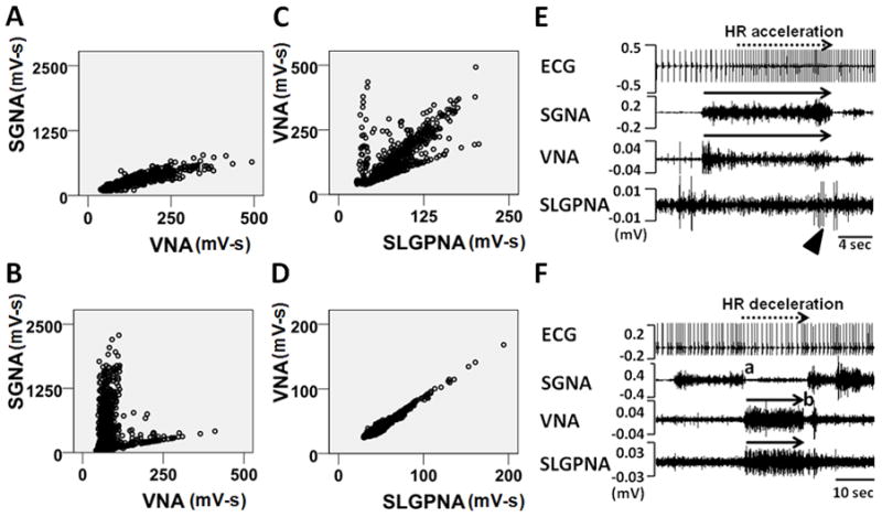
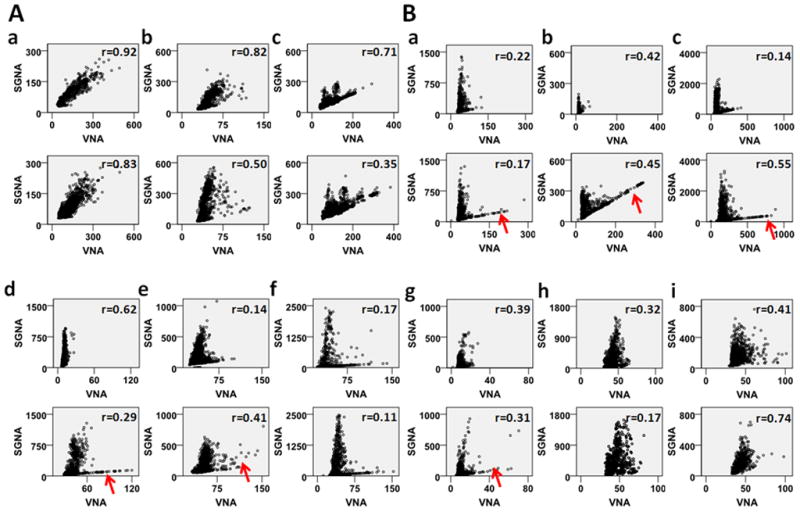
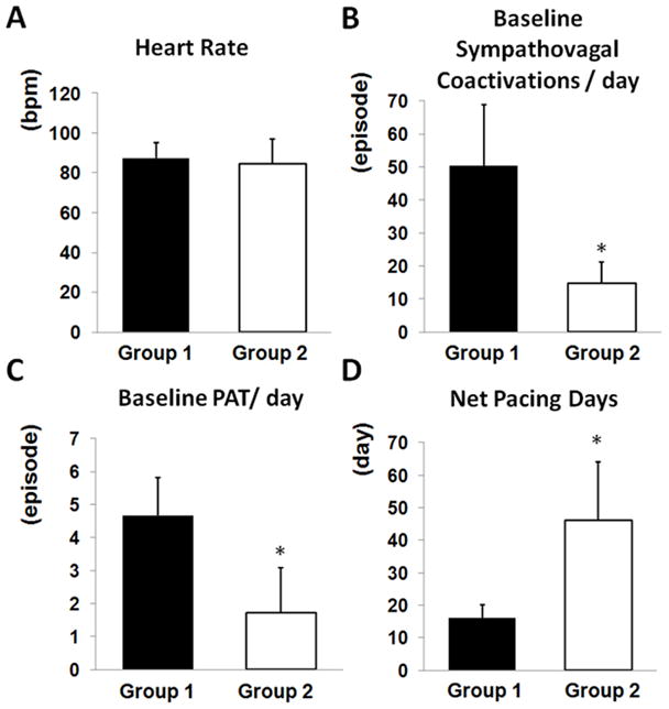
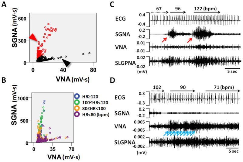
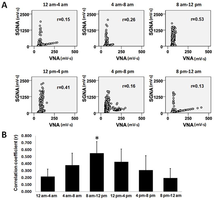
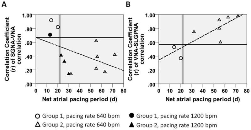
Results: At baseline (before pacing), 1-minute integrated nerve activity between SGNA and VNA demonstrated either a single linear relationship with excellent correlation (group 1, N = 3, r = 0.816 ± 0.105) or nonlinear relationships with poor correlation (group 2, N = 9, r = 0.316 ± 0.162, P <.05 vs group 1). Group 1 dogs had higher VNA (97.0 ± 11.5 mV-s) compared to group 2 (33.4 ± 21.7 mV-s, P <.001). Group 1 dogs had more frequent sympathovagal co-activation episodes than did group 2 (50 ± 19 per day vs 15 ± 6 per day, P <.05) and more paroxysmal atrial tachycardia (PAT; 5 ± 1 per day vs 2 ± 1 per day, P <.05) at baseline. Sustained AF occurred after 16 ± 4 days (range 13-20 days) of pacing in group 1 and after 46 ± 18 days (range 23-72 days) of pacing in group 2 (P <.05). In the week before development of sustained AF, VNA of group 2 dogs was significantly increased compared to baseline (P <.05).

Conclusion: Ambulatory dogs with good linear sympathovagal correlation and higher vagal tone at baseline have more PAT episodes at baseline and faster induction of sustained AF by rapid pacing. Rapid atrial pacing increased the VNA of the remaining dogs before induction of sustained AF.


Figures

Figure 1
Figure 1
Patterns of autonomic interactions. A, Representative SGNA-VNA scatter plot of a Group 1 dog. Each dot represents an SGNA-VNA pair of nerve activity integrated over 1-min. The entire plot has 1440 data points to cover a 24-hr period. B, Representative SGNA-VNA scatter plot from a Group 2 dog. C, Representative VNA-SLGPNA scatter plot from a Group 1 dog. D, Representative VNA-SLGPNA scatter plot from a Group 2 dog. E, An example of simultaneous sympathovagal co-activation (black arrows) observed in a Group 1 dog that led to heart rate acceleration. Arrowhead shows independent SLGPNA. F, An example of recording from a Group 2 dog showing that simultaneously increased VNA and SLGPNA (black arrows) resulted in heart rate deceleration. ECG, electrocardiogram; SGNA, stellate ganglion nerve activity; VNA, vagal nerve activity; SLGPNA, superior left ganglionated plexi nerve activity.
Figure 2
Figure 2
Sympathovagal correlations of all dogs studied. Panels A and B show dogs with excellent correlations (Group 1) and poor correlations (Group 2), respectively. The dogs of Group 2 show “L-shaped” correlation. However, because this designation is subjective, it is not used to determine the grouping of the dogs. The upper panels show the baseline correlation, while the lower panels show correlation in the week before the development of sustained AF. We used the same scales of the ordinate and abscissa within each dog to facilitate the comparison. The red arrows point to apparent increase of lower (vagal) arm of the correlation in six Group 2 dogs prior to the onset of sustained AF. All units are mV-s.
Figure 3
Figure 3
Comparing electrophysiologic characteristics between two groups of dogs. A, Baseline heart rate did not differ significantly between two groups. B, Group 1 dogs had more episodes of prolonged (≥5 sec) sympathovagal coactivations per day at baseline. C, Group 1 dogs had more episodes of PAT per day at baseline. D, Group 1 dogs required much fewer days of pacing than Group 2 dogs before developing sustained AF. *P<0.05.
Figure 4
Figure 4
The physiology of L-shaped sympathovagal correlation. A, An example of a L-shaped sympathovagal plot. The upper and lower arms are coded with red and black circles, respectively. B, L-shaped sympathovagal plot from another dog. Each dot is labeled with different colors according to the average heart rate within the 1-min interval represented by each dot. The upper arm shows progressively increasing SGNA is associated with a progressively higher heart rate. The lower arm shows that increasing VNA is associated with increasing SGNA and heart rate. However, because all dots in the lower arm are associated with a heart rate of < 100 bpm, VNA has exerted a dominant effect on heart rate in these time points. C, Actual recordings at the red arrowhead in Panel A. Vigorous SGNA (pointed by a red arrow) caused heart rate acceleration. D, Actual recordings at the black arrowhead in Panel A. Gradually increasing VNA (pointed by blue arrows) led to progressive heart rate deceleration.
Figure 5
Figure 5
Circadian variations of the sympathovagal relationships. A shows the sympathovagal plots from one representative dog according to the time periods within 24 hrs. The Pearson’s correlation coefficients (r) are also shown on the corresponding plots. B, The circadian variations of the correlation coefficients of all dogs studied. *P<0.001 compared to 12 am–4 am.
Figure 6
Figure 6
The relationship between autonomic interactions at baseline and the susceptibility to pacing induced sustained AF. A, Better SGNA-VNA correlation is associated with shorter periods of atrial tachypacing needed for induction of sustained AF. The Group 1 dogs (circles) are exclusively in the left upper quadrant of the scatter plot whereas the Group 2 dogs (triangles) are entirely in the right lower one. Dogs paced at 1200 bpm are labeled with filled circles/triangles while those paced at 640 bpm are labeled with unfilled circles/triangles. B, Poorer VNA-SLGPNA correlation is associated with shorter periods of atrial rapid pacing.

Comment in

References

    1. Kerr CR, Humphries KH, Talajic M, et al. Progression to chronic atrial fibrillation after the initial diagnosis of paroxysmal atrial fibrillation: results from the Canadian Registry of Atrial Fibrillation. Am Heart J. 2005;149:489–496. - PubMed
    1. Jahangir A, Lee V, Friedman PA, et al. Long-term progression and outcomes with aging in patients with lone atrial fibrillation: a 30-year follow-up study. Circulation. 2007;115:3050–3056. - PubMed
    1. Tan AY, Zhou S, Ogawa M, et al. Neural mechanisms of paroxysmal atrial fibrillation and paroxysmal atrial tachycardia in ambulatory canines. Circulation. 2008;118:916–925. - PMC - PubMed
    1. Choi E-K, Shen MJ, Han S, et al. Intrinsic cardiac nerve activity and paroxysmal atrial tachyarrhythmia in ambulatory dogs. Circulation. 2010;121:2615–2623. - PMC - PubMed
    1. Wijffels MC, Kirchhof CJ, Dorland R, Allessie MA. Atrial fibrillation begets atrial fibrillation. A study in awake chronically instrumented goats. Circulation. 1995;92:1954–1968. - PubMed

Publication types