

Bidirectional ventricular tachycardia: ping pong in the His-Purkinje system
- PMID: 21118730
- PMCID: PMC3066157
- DOI: 10.1016/j.hrthm.2010.11.038
Bidirectional ventricular tachycardia: ping pong in the His-Purkinje system
Abstract
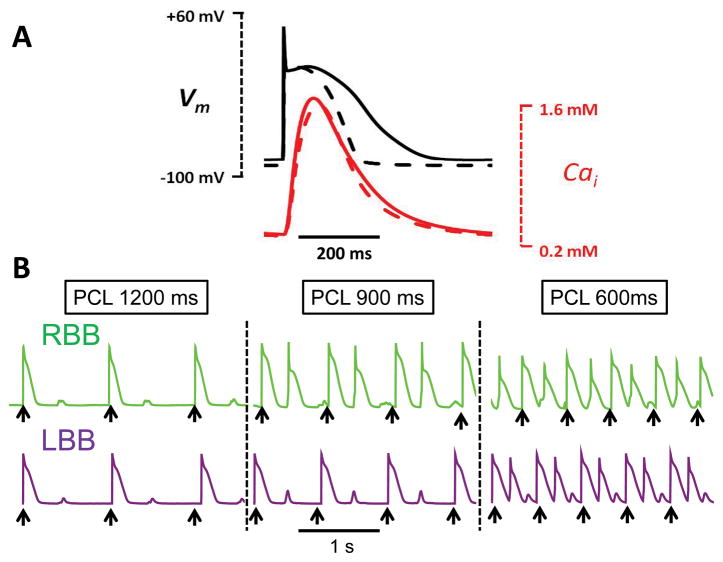
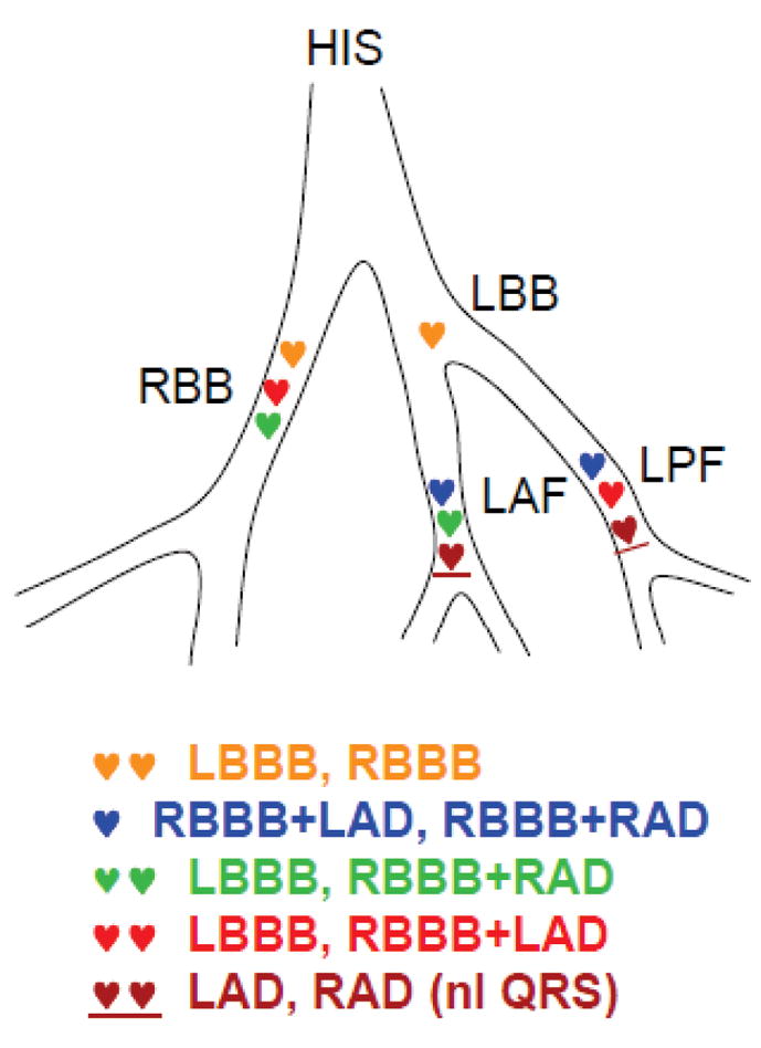
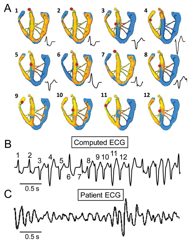
Background: Bidirectional ventricular tachycardia (BVT), which is characterized by an alternating beat-to-beat ECG QRS axis, is a rare but intriguing arrhythmia associated with digitalis toxicity, familial catecholaminergic polymorphic ventricular tachycardia (CPVT), and several other conditions that predispose cardiac myocytes to delayed afterdepolarizations (DADs) and triggered activity. Evidence from human and animal studies attributes BVT to alternating ectopic foci originating from the distal His-Purkinje system in the left and/or right ventricle, respectively.
Objective: The purpose of this study was to evaluate a simple "ping pong" model of reciprocating bigeminy to explain BVT.
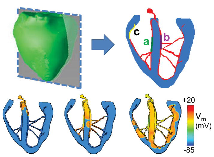
Methods: We constructed a two-dimensional anatomic model of the rabbit ventricles with a simplified His-Purkinje system, in which different sites in the His-Purkinje system had different heart rate thresholds for DAD-induced bigeminy.
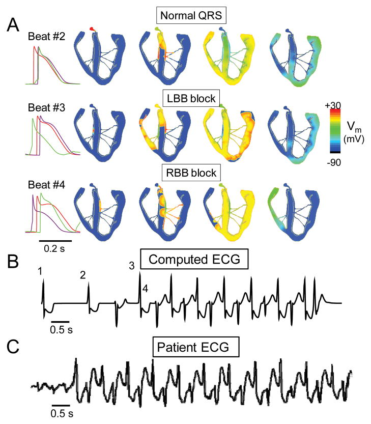
Results: When the heart rate exceeded the threshold for bigeminy at the first site in the His-Purkinje system, ventricular bigeminy developed, causing the heart rate to accelerate and exceed the threshold for bigeminy at the second site. Thus, the triggered beat from the first site induced a triggered beat from the second site. The triggered beat from the second site next reciprocated by inducing a triggered beat from the first site, and so forth. Bigeminy from two sites produced BVT, and that from three or more sites produced polymorphic VT.
Conclusion: This "ping pong" mechanism of reciprocating bigeminy readily produces the characteristic ECG pattern of BVT and its degeneration to polymorphic VT if additional sites develop bigeminy.
Copyright © 2011 Heart Rhythm Society. Published by Elsevier Inc. All rights reserved.
Conflict of interest statement
Figures
Comment in
-
Serving up the ping-pong mechanisms for biventricular ventricular tachycardia.Heart Rhythm. 2011 Apr;8(4):606-7. doi: 10.1016/j.hrthm.2011.02.031. Epub 2011 Feb 23. Heart Rhythm. 2011. PMID: 21354333 No abstract available.
References
-
- Schwensen C. Ventricular Tachycardia as the result of the administration of digitalis. Heart. 1922;9:199–204.
-
- Valent S, Kelly P. Digoxin-Induced Bidirectional Ventricular Tachycardia. N Engl J Med. 1997;336(8):550. - PubMed
-
- Kummer JL, Nair R, Krishnan SC. Bidirectional Ventricular Tachycardia Caused by Digitalis Toxicity. Circulation. 2006;113(7):e156–157. - PubMed
-
- Menduiña MJ, Candel JM, Alaminos P, et al. Bidirectional Ventricular Tachycardia Due to Digitalis Poisoning. Revista Espanola de Cardiologia. 2005;58(8):991–993. - PubMed
Publication types
MeSH terms
Supplementary concepts
Grants and funding
LinkOut - more resources
Full Text Sources
