. 2011 Jan 15;22(2):245-55.
doi: 10.1091/mbc.E10-07-0633. Epub 2010 Nov 30.

H4 replication-dependent diacetylation and Hat1 promote S-phase chromatin assembly in vivo

Affiliations

H4 replication-dependent diacetylation and Hat1 promote S-phase chromatin assembly in vivo

Aïda Ejlassi-Lassallette et al. Mol Biol Cell. .

Abstract

While specific posttranslational modification patterns within the H3 and H4 tail domains are associated with the S-phase, their actual functions in replication-dependent chromatin assembly have not yet been defined. Here we used incorporation of trace amounts of recombinant proteins into naturally synchronous macroplasmodia of Physarum polycephalum to examine the function of H3 and H4 tail domains in replication-coupled chromatin assembly. We found that the H3/H4 complex lacking the H4 tail domain was not efficiently recovered in nuclei, whereas depletion of the H3 tail domain did not impede nuclear import but chromatin assembly failed. Furthermore, our results revealed that the proper pattern of acetylation on the H4 tail domain is required for nuclear import and chromatin assembly. This is most likely due to binding of Hat1, as coimmunoprecipitation experiments showed Hat1 associated with predeposition histones in the cytoplasm and with replicating chromatin. These results suggest that the type B histone acetyltransferase assists in shuttling the H3/H4 complex from cytoplasm to the replication forks.


Figures

FIGURE 1:
FIGURE 1:
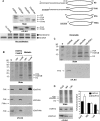
Exogenous FLAG-tagged H3/H4 histones incorporated into nuclei and assembled in chromatin in a replication-coupled manner. (A) Experimental scheme: Macroplasmodium is cut in two halves in early S-Phase (S-phase lasts 3 h in Physarum). One half is treated with a solution containing purified histones, and the other half is used as control. The cell fragments are harvested after 1 h treatment and analyzed. (B) Exogenous histones H3/H4 are incorporated into nuclei. Following cell fractionation, samples (total, cytoplasm, and nuclei) without exogenous histones (control) and with exogenous histones (H3/FH4) are resolved in SDS–PAGE and analyzed by Western blotting with anti-FLAG antibody. (C) FLAG-tagged exogenous H3/H4 colocalized with DAPI-stained Physarum nuclei. Cell explants were squashed, fixed, and stained with anti-FLAG antibody. Slides were then observed by fluorescence microscopy. (D) Exogenous histone H3/H4 complex is assembled in chromatin. Nuclear fraction from (B) was used for preparing soluble chromatin with hydroxyapatite beads and high-salt wash. Chromatin-bound fractions are then analyzed by SDS–PAGE and Western blotting. (E) Accumulation of exogenous H3/H4 into nuclei depends upon replication activity. Plasmodium fragments are treated with exogenous histones in the presence and absence of hydroxyurea (HU). Cell fractions (total, cytoplasm, and nuclei) are analyzed similarly to (B). The percentages below the blot correspond to the quantification of overexposed blots (unpublished data) and represent the percentage of histone in cytoplasmic fractions relative to histone incorporated into the macroplasmodia. (F) HU treatment blocks nuclear import of exogenous histones. Fluorescence microscopy observations of cell explants treated as in (C). Control treated with exogenous H3/H4 in the absence of HU revealed a colocalization of FLAG signal with DAPI-stained nuclei. HU-treated cells concomitantly with exogenous histone incorporation showed the absence of accumulation into nuclei, but cytoplasmic aggregates are observed (arrowheads). (G) Blocking replication prevents chromatin assembly. Nuclear fractions from (E) were analyzed as in (D).
FIGURE 2:
FIGURE 2:
Tail domain regions of H3 and H4 exhibit distinct functions in cellular localization and chromatin assembly requirement. (A) Histone mutants of H3 and H4 were used to determine the function of the histone tail domains during S-phase. Schemes represent the histones that are expressed in Escherichia coli. The underlined and italicized sequences at the amino termini of FH4 and FH4n correspond to FLAG sequences that are used throughout our experiments. The histone complexes have been assessed by SDS–PAGE and Western blotting, and their ability to form nucleosome in vitro has been evaluated relative to purified chicken erythrocyte H3/H4 (Ch H3/H4). (B) Cellular localization of exogenous H3/H4 complexes. Exogenous histone incorporations and cell fractionation are carried out similarly to Figure 1. Each mutant complex is incorporated in half macroplasmodium, and the other half is treated with full-length histones and used as control. Shown on the top is a representative SDS–PAGE of cell fractionations. The bottom panels correspond to blots obtained with the different mutant complexes, H3n/FH4, H3/FH4n, and H3n/FH4n, respectively. (C) Chromatin analyses revealed the requirement of H3 and H4 tail domains in replication-coupled chromatin assembly. Chromatin is prepared from different nuclear fractions and analyzed by SDS–PAGE and Western blotting. Note that the absence of immunodetection with H3n/FH4 indicates that the exogenous complex is not disrupted on incorporation and does not exchange with Physarum histones. (D) Depletion of the H3 amino-tail domain affects stability of exogenous complexes in nuclei. H3/FH4 and H3n/FH4 are incorporated into Physarum at the beginning of S-phase, and plasmodium fragments are harvested after 1 h (early), 2 h (mid), and 3 h (late), respectively. Nuclei are isolated and analyzed by SDS–PAGE and Western blotting, wherein a constant amount of DNA is loaded. The graph represents the quantitation of three different experiments in which 100% was arbitrarily affected to full-length exogenous histones in early S-phase (mean ± SEM).
FIGURE 3.
FIGURE 3.
Histone H4 acetylation is required for nuclear import and facilitates chromatin assembly. (A) Mutations of lysine residues 5 and 12 of H4. Scheme represents the substitutions of lysines 5 and 12 to glutamine (Q) to mimic acetylation and to arginine (R) to prevent acetylation. (B) Cellular localization of the different histone complexes. Exogenous histones were incorporated for 1 h in early S-phase, similarly to Figure 1, and cells were fractionated before electrophoretic analyses of the fractions. (C) Chromatin assembly analyses of exogenous mutant complexes. Chromatin is prepared from nuclear fractions obtained after exogenous histone complex treatment, H3/FH4, H3/FH4-Q5/12, and H3/FH4-R5/12, respectively.
FIGURE 4:
FIGURE 4:
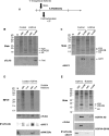
Hat1 binding to H3/H4 in cytoplasm is required for the delocalization of the tertiary complex (Hat1–H3/H4) into nuclei. (A) Experimental scheme of the incorporation of exogenous histones. H3/FH4 complex is incorporated in Physarum plasmodium in early S-phase for a short period of 10 min. Cells are then harvested and fractionated before carrying out immunoprecipitation (IP) experiments. Plasmodium fragment untreated with exogenous histones was used as control. (B) Cellular localization of exogenous H3/FH4. Following cell fractionations, cytoplasmic (cyto) and nuclear fractions (nuclei), respectively, were analyzed by SDS–PAGE and Western blotting. Note that even after the 10-min treatment with exogenous histones, H3/FH4 complex is recovered into nuclear fraction. (C) Cellular localization of HAT1. Cytoplasmic (cyto) and nuclear (nuclei) fractions from control and H3/FH4-treated cells are analyzed by SDS–PAGE and Western blotting. In control and H3/FH4-treated fragments, anti-HAT1 antibodies recognize a band of ∼50 kDa corresponding to the molecular mass of HAT1. (D) IP of FLAG-containing complexes. Soluble fractions from cytoplasmic and nuclear fractions are prepared from control and exogenous histone-treated cells (Input) and used for immunoprecipitating FLAG-containing complexes. Immunoprecipitated proteins are analyzed by Western blotting using specific antibodies, anti-FLAG (αFLAG), anti-HAT1 (αHAT1), and anti-H4K12Acetyl (αH4K12Ac), respectively. The input corresponds to 1:2000 of the IP. Note that analyses with anti-H4K8Acetyl and anti-H4K5Acetyl did not reveal H4 acetylation (unpublished data). (E) Preventing Hat1 binding to H4 reduces the accumulation of H3/H4 in nuclei. The complexes H3/FH4 (control), H3/FH4-4Q (K5, K8, K12, and K16 substituted to Q), and H3/FH4-Q8/16 (K8 and K16 substituted to Q) were incorporated in Physarum cells for 1 h in early S-phase. Following cell fractionations, the cytoplasmic (cyto) and the nuclear (nuclei) fractions were resolved in SDS–PAGE and analyzed by Western blotting. IP of FLAG-containing complexes in soluble cytoplasmic and soluble nuclear fractions was carried out as in (D) on cells treated with H3/FH4 and H3/FH4-Q8/16, and the presence of Hat1 was assessed by Western blotting.
FIGURE 5:
FIGURE 5:
Purification and analysis of the replication complex. (A) Experimental scheme of the procedure of purification of replication complex. (B) Silver-stained SDS–PAGE of input (Input), immunoprecipitated material (IP), and immunoprecipitated material in the presence of competitor bacterial DNA (Compet), respectively. The molecular mass markers are 250, 150, 100, 75, 50, 37, 25, 20, 15, and 10 kDa, respectively. (C) Analysis of HAT1 in replication complex. Antibodies to HAT1 are used to reveal Western blot of untreated cell fractions (Untreated), bound (IP) and unbound (UN) fractions from chromatin untreated with BrdU (No BrdU), BrdU immunoprecipitated chromatin (IP BrdU), and PCNA immunoprecipitated chromatin in conditions identical to IP BrdU (IP PCNA), respectively. Densitometric profiles of immunostained HAT1 band are shown.
FIGURE 6:
FIGURE 6:
Model of the role of HAT1 in the supply of H3/H4 dimer at the replication fork. During S-phase, the newly synthesized H3/H4 dimer associates with HAT1 in the cytoplasm, where H4 is acetylated at least at lysine 12. Concomitantly to DNA replication, the complex composed of HAT1 and H3/H4 is transferred into nuclei and localizes in the vicinity of replicating chromatin for supplying H3/H4 to chromatin assembly factors.

References

    1. Ahmad A, Takami Y, Nakayama T. Distinct regions of the chicken p46 polypeptide are required for its in vitro interaction with histones H2B and H4 and histone acetyltransferase-1. Biochem Biophys Res Commun. 2000;279:95–102. - PubMed
    1. Ai X, Parthun MR. The nuclear Hat1p/Hat2p complex: a molecular link between type B histone acetyltransferases and chromatin assembly. Mol Cell. 2004;14:195–205. - PubMed
    1. Almouzni G, Mechali M. Assembly of spaced chromatin promoted by DNA synthesis in extracts from Xenopus eggs. EMBO J. 1988;7:665–672. - PMC - PubMed
    1. Annunziato AT, Hansen JC. Role of histone acetylation in the assembly and modulation of chromatin structures. Gene Expr. 2000;9:37–61. - PMC - PubMed
    1. Barman HK, Takami Y, Ono T, Nishijima H, Sanematsu F, Shibahara K, Nakayama T. Histone acetyltransferase 1 is dispensable for replication-coupled chromatin assembly but contributes to recover DNA damages created following replication blockage in vertebrate cells. Biochem Biophys Res Commun. 2006;345:1547–1557. - PubMed

Publication types

MeSH terms

Substances

LinkOut - more resources