Review
. 2010 Dec;74(4):504-28.
doi: 10.1128/MMBR.00021-10.

FtsZ in bacterial cytokinesis: cytoskeleton and force generator all in one

Affiliations
Review

FtsZ in bacterial cytokinesis: cytoskeleton and force generator all in one

Harold P Erickson et al. Microbiol Mol Biol Rev. 2010 Dec.

Abstract

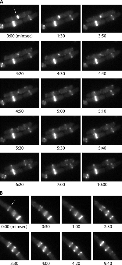
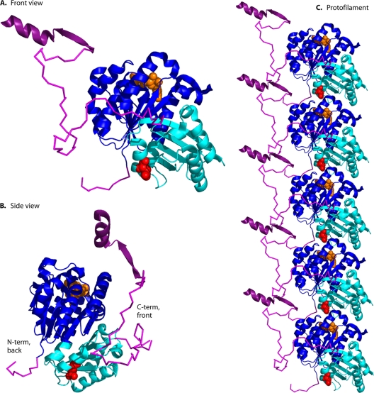
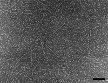
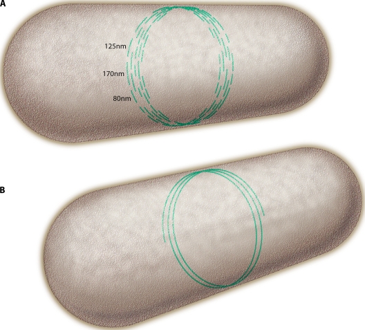
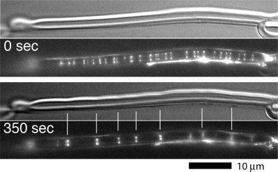
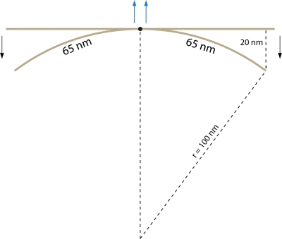
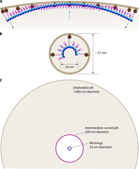
FtsZ, a bacterial homolog of tubulin, is well established as forming the cytoskeletal framework for the cytokinetic ring. Recent work has shown that purified FtsZ, in the absence of any other division proteins, can assemble Z rings when incorporated inside tubular liposomes. Moreover, these artificial Z rings can generate a constriction force, demonstrating that FtsZ is its own force generator. Here we review light microscope observations of how Z rings assemble in bacteria. Assembly begins with long-pitch helices that condense into the Z ring. Once formed, the Z ring can transition to short-pitch helices that are suggestive of its structure. FtsZ assembles in vitro into short protofilaments that are ∼30 subunits long. We present models for how these protofilaments might be further assembled into the Z ring. We discuss recent experiments on assembly dynamics of FtsZ in vitro, with particular attention to how two regulatory proteins, SulA and MinC, inhibit assembly. Recent efforts to develop antibacterial drugs that target FtsZ are reviewed. Finally, we discuss evidence of how FtsZ generates a constriction force: by protofilament bending into a curved conformation.


Figures

FIG. 1.
FIG. 1.
Time-lapse observation of Z rings in E. coli, using FtsZ-GFP as a dilute label, expressed at about one-third the level of genomic FtsZ. The cells were induced to produce FtsZ-GFP for about 1 h and then immobilized on an agar pad for time-lapse observation at 37°C. (A) Three cells. We will ignore the two on the bottom (with a bright Z ring [left] and a dim Z ring [right]) and focus on the upper one (arrow), which is undergoing division. The constriction of the Z ring, its concurrent disassembly, and the assembly of new Z rings in the daughter cells are described in the text. (B) A cell with three Z rings (perhaps induced by excessive production of FtsZ-GFP). The upper Z ring (arrow) alternately opens into a short-pitch helix and collapses into an apparent circle. Frames from both panels A and B are taken from Movie S1 in the supplemental material.
FIG. 2.
FIG. 2.
(A) Structure of the FtsZ subunit. The globular domain, shown in cartoon format, comprises two subdomains colored blue (N-terminal) and cyan (C-terminal). This is from the X-ray structure of P. aeruginosa FtsZ, PDB 1OFU (32). The GDP is shown in orange space fill, and the synergy loop amino acid D212 (E. coli numbering) is in red. This view corresponds to that of a tubulin subunit seen from the outside of a microtubule, and is designated the “front view.” A 10-amino-acid segment on the N terminus and a 50-amino-acid segment on the C terminus are shown in magenta, each modeled as flexible peptides. Shown in dark purple are the extended beta strand and alpha helix formed by the C-terminal 17-amino-acid peptide when bound to ZipA (from PDB 1F47 [121]). The model was constructed using the program PyMol (39). (B) The FtsZ subunit viewed from the side. This shows that the C-terminal peptide emerges from the front face and the N-terminal peptide from the back face, ∼180 degrees away. (C) A protofilament is assembled by stacking subunits on top of each other so that the D212 of the upper subunit is just above the GDP of the one below.
FIG. 3.
FIG. 3.
Electron micrograph of negatively stained protofilaments assembled in vitro from E. coli FtsZ (1 μM FtsZ, 50 mM MES [morpholineethanesulfonic acid] [pH 6.5], 100 mM KAc, 5 mM MgAc, 1 mM GTP). The bar is 100 nm. This specimen was prepared on a carbon film treated with UV light and ozone to render it hydrophilic (24). With these carbon films we obtain protofilaments at lower FtsZ concentrations, and they are longer than those previously reported. The protofilaments here are mostly straight, but some show a tendency to curve.
FIG. 4.
FIG. 4.
(A) A model for how short protofilaments might be arranged to make the Z ring. The average 125-nm length is much shorter than the 3,000-nm circumference, so protofilaments would be arranged in a staggered overlap. (B) An alternative structure where the short protofilaments are proposed to anneal into one or a few long protofilaments.
FIG. 5.
FIG. 5.
Z rings assembled in tubular liposomes from purified, membrane-targeted FtsZ. The upper frame at the two times uses Nomarski optics to show the profile of the liposome. The lower panel shows FtsZ localized by yellow fluorescent protein (YFP) fluorescence. At time zero (actually about 10 min after specimen preparation), there are many dim Z rings. After 350 s, the dim Z rings have coalesced into fewer bright ones, and these are generating constrictions in the liposome. The full sequence may be seen in Movie S2 in the supplemental material. (This figure is reprinted and Movie S2 is reproduced from reference with permission from AAAS.)
FIG. 6.
FIG. 6.
Scale model of protofilament bending. A 130-nm-long protofilament (solid line) is shown fixed at the center (dot) in the straight and the intermediate curved conformations. The two 65-nm halves behave as individual cantilevered beams as the protofilament bends to the radius of the intermediate curved conformation. Arrows show the direction of force (lighter central arrows represent the opposite force that would necessarily push back against the membrane if the protofilament were free).
FIG. 7.
FIG. 7.
FtsZ on the outside of liposomes produces distortions. (A) Normal membrane-targeted FtsZ, with the membrane tether on the C terminus, forms concave depressions. (B) When the tether is switched to the N terminus, it forms convex protrusions. (C) Diagram showing the curvature and two sides for attaching the membrane targeting sequence (MTS). (Reprinted from reference with permission from Macmillan Publishers Ltd.)
FIG. 8.
FIG. 8.
Asymmetric division of isolated cyanelle plastids. (A) Three cyanelles imaged by scanning EM, showing furrows progressing from shallow (arrow) to deep (double arrowhead). (B to D) Increasingly deep furrows correspond to increasing length of the FtsZ arc. Row 1 shows Nomarski images, row 2 shows the cytoplasm imaged by autofluorescence of chlorophyll and phycobilin, row 3 shows immunofluorescence staining of FtsZ, and row 4 shows overlap of rows 2 and 3. (Reprinted from reference with kind permission from Springer Science+Business Media.)
FIG. 9.
FIG. 9.
Bending protofilaments can only constrict so far. (A) An FtsZ protofilament is shown tethered to the membrane of an undivided cell. The blue FtsZ subunits are connected into a protofilament that has a preferred bend. About one out of five FtsZ proteins are shown tethered to an FtsA, indicated by an orange circle. An amphipathic helix extends from FtsA into the membrane bilayer. The FtsZ tether is shown extended to ∼10 nm at the ends of the protofilament, where the bending toward the cell center is maximal. Near the middle of the protofilament it is pushing outward on the membrane, and the tethers are compressed. (B) When fully in the curved conformation, the 24-nm miniring plus an ∼10-nm tether plus FtsA could constrict the membrane to about a 57-nm outside diameter. (C) The diameters of the undivided cell, the intermediate curved conformation, and the miniring are compared.

References

    1. Aarsman, M. E., A. Piette, C. Fraipont, T. M. Vinkenvleugel, M. Nguyen-Disteche, and T. den Blaauwen. 2005. Maturation of the Escherichia coli divisome occurs in two steps. Mol. Microbiol. 55:1631-1645. - PubMed
    1. Adams, D. W., and J. Errington. 2009. Bacterial cell division: assembly, maintenance and disassembly of the Z ring. Nat. Rev. Microbiol. 7:642-653. - PubMed
    1. Addinall, S. G., E. F. Bi, and J. Lutkenhaus. 1996. FtsZ ring formation in fts mutants. J. Bacteriol. 178:3877-3884. - PMC - PubMed
    1. Addinall, S. G., C. Cao, and J. Lutkenhaus. 1997. Temperature shift experiments with ftsZ84(Ts) strain reveal rapid dynamics of FtsZ localization and indicate that the Z ring is required throughout septation and cannot reoccupy division sites once constriction has initiated. J. Bacteriol. 179:4277-4284. - PMC - PubMed
    1. Addinall, S. G., and J. Lutkenhaus. 1996. FtsZ-spirals and -arcs determine the shape of the invaginating septa in some mutants of Escherichia coli. Mol. Microbiol. 22:231-237. - PubMed

Publication types

MeSH terms

LinkOut - more resources