Review
. 2010 Dec;74(4):552-69.
doi: 10.1128/MMBR.00024-10.

Cell biology of the trypanosome genome

Affiliations
Review

Cell biology of the trypanosome genome

Jan-Peter Daniels et al. Microbiol Mol Biol Rev. 2010 Dec.

Abstract

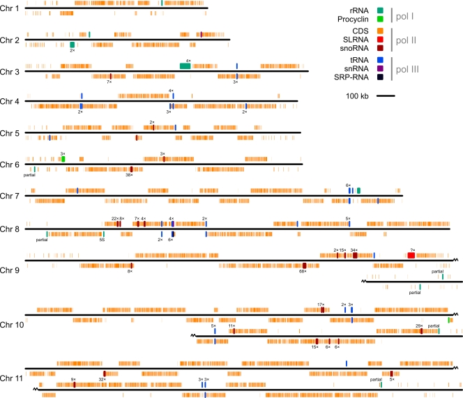
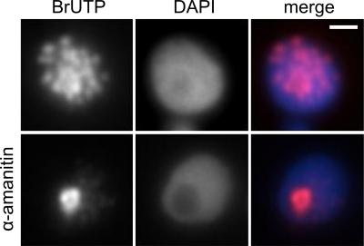
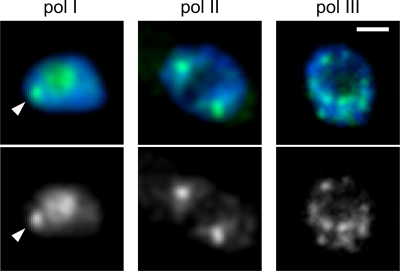
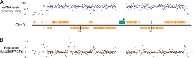
Trypanosomes are a group of protozoan eukaryotes, many of which are major parasites of humans and livestock. The genomes of trypanosomes and their modes of gene expression differ in several important aspects from those of other eukaryotic model organisms. Protein-coding genes are organized in large directional gene clusters on a genome-wide scale, and their polycistronic transcription is not generally regulated at initiation. Transcripts from these polycistrons are processed by global trans-splicing of pre-mRNA. Furthermore, in African trypanosomes, some protein-coding genes are transcribed by a multifunctional RNA polymerase I from a specialized extranucleolar compartment. The primary DNA sequence of the trypanosome genomes and their cellular organization have usually been treated as separate entities. However, it is becoming increasingly clear that in order to understand how a genome functions in a living cell, we will need to unravel how the one-dimensional genomic sequence and its trans-acting factors are arranged in the three-dimensional space of the eukaryotic nucleus. Understanding this cell biology of the genome will be crucial if we are to elucidate the genetic control mechanisms of parasitism. Here, we integrate the concepts of nuclear architecture, deduced largely from studies of yeast and mammalian nuclei, with recent developments in our knowledge of the trypanosome genome, gene expression, and nuclear organization. We also compare this nuclear organization to those in other systems in order to shed light on the evolution of nuclear architecture in eukaryotes.


Figures

FIG. 1.
FIG. 1.
Map of the T. brucei genome showing the organization of genes according to their class and transcribing polymerase. The map is based on the v4 annotation available at www.genedb.org. Colored bars indicate the positions and lengths of different genetic elements relative to the chromosome backbone (black line). Bars above the lines indicate transcription toward the right; bars below the lines indicate transcription toward the left. When a number of a similar elements are present in close proximity in the genome, this is indicated by n× next to bar (where n is the number of elements). Noncoding RNA genes have been given a minimum bar length to facilitate visualization. Only the largest assembled contig is represented for each chromosome. Chromosomes 9 to 11 have been split across two lines.
FIG. 2.
FIG. 2.
Compartmentalization of transcription in the trypanosome nucleus. Images show the incorporation of BrUTP into saponin-permeabilized nuclei in the absence (top row) or presence (bottom row) of α-amanitin, which inhibits transcription by pol II and pol III. A stain of the nuclear DNA (DAPI) is also shown. Images courtesy of Eva Gluenz (University of Oxford, United Kingdom).
FIG. 3.
FIG. 3.
Distribution of subunits of pol I, pol II and pol III in the T. brucei nucleus. (pol I) Native fluorescence of RPA2, the second-largest subunit of pol I, N-terminally tagged in bloodstream-form cells with the TY epitope and yellow fluorescent protein (YFP). Cells were fixed with 3% formaldehyde in phosphate-buffered saline (PBS). (pol II) Largest subunit of pol II detected by immunofluorescence with an anti-RPB1 polyclonal antibody (a gift from V. Bellofatto, New Jersey Medical School). Cell were fixed in 4% formaldehyde and 5% acetic acid in PBS. (pol III) Native fluorescence of C-terminally YFP-TY-tagged RPC1, largest subunit of pol III. Cells were fixed in 3% formaldehyde in PBS. In the overlay images, the YFP signal is pseudocolored in green and the DAPI signal in blue. Bar, 1 μm.
FIG. 4.
FIG. 4.
General lack of gene expression regulation at the level of pol II transcription units in trypanosomes. A map of T. brucei chromosome 3 is shown with measurements of total mRNA levels (in cultured bloodstream-form cells) (A) and the fold change in mRNA levels between cultured bloodstream- and procyclic-form cells (B). The map is based on the v4 genome annotation available from GeneDB (www.genedb.org). mRNA measurements are taken from a study by Jensen et al. (86) which has been made publicly available under accession number GSE18049 in the GEO database (www.ncbi.nlm.nih.gov/geo/).
FIG. 5.
FIG. 5.
Ultrastructural compartmentalization of the trypanosome nucleolus. The image shows a transmission electron micrograph of a thin section though a bloodstream-form T. brucei nucleus. The most obvious nuclear compartment is the nucleolus (N) in the center of the micrograph, but there is also a clear distinction between electron-lucent (1, “euchromatin”) and electron-dense (2, “heterochromatin”) regions in the nucleoplasm. The nucleus is surrounded by the nuclear envelop (ne), in which can be seen nuclear pores (np) which are always juxtaposed with electron-lucent nucleoplasm. The sample was prepared by high-pressure freezing, followed by freeze substitution with 1% glutaraldehyde and 2% uranyl acetate in acetone and then 2% osmium tetroxide and 2% uranyl acetate in acetone and embedding in resin. Image courtesy of Catarina Gadelha (University of Cambridge, United Kingdom).
FIG. 6.
FIG. 6.
Concentration of nuclear proteins in the nucleolar periphery. Procyclic cells fixed in 3% formaldehyde are shown. N-terminally TY-YFP-tagged TFIIS2-1 was detected with an anti-TY antibody (BB2, pseudocolored in green) and Nopp140-like protein with an anti-Nopp140-like antibody (pseudocolored in red). DAPI is pseudocolored in blue. Bar, 1 μm. The two proteins concentrate in different parts of the nucleolar periphery, resulting in an incomplete colocalization of signals.

References

    1. Abanades, D. R., L. Ramirez, S. Iborra, K. Soteriadou, V. M. Gonzalez, P. Bonay, C. Alonso, and M. Soto. 2009. Key role of the 3′ untranslated region in the cell cycle regulated expression of the Leishmania infantum histone H2A genes: minor synergistic effect of the 5′ untranslated region. BMC Mol. Biol. 10:48. - PMC - PubMed
    1. Alsford, S., B. Wickstead, K. Ersfeld, and K. Gull. 2001. Diversity and dynamics of the minichromosomal karyotype in Trypanosoma brucei. Mol. Biochem. Parasitol. 113:79-88. - PubMed
    1. Amiguet-Vercher, A., D. Pérez-Morga, A. Pays, P. Poelvoorde, H. Van Xong, P. Tebabi, L. Vanhamme, and E. Pays. 2004. Loss of the mono-allelic control of the VSG expression sites during the development of Trypanosoma brucei in the bloodstream. Mol. Microbiol. 51:1577-1588. - PubMed
    1. Archer, S. K., V. Luu, R. A. de Queiroz, S. Brems, and C. Clayton. 2009. Trypanosoma brucei PUF9 regulates mRNAs for proteins involved in replicative processes over the cell cycle. PLoS Pathog. 5:e1000565. - PMC - PubMed
    1. Avliyakulov, N. K., J. C. Hines, and D. S. Ray. 2003. Sequence elements in both the intergenic space and the 3′ untranslated region of the Crithidia fasciculata KAP3 gene are required for cell cycle regulation of KAP3 mRNA. Eukaryot. Cell 2:671-677. - PMC - PubMed

Publication types

LinkOut - more resources