. 2010 Nov;22(11):3791-803.
doi: 10.1105/tpc.110.078378. Epub 2010 Nov 30.

Meiotic progression in Arabidopsis is governed by complex regulatory interactions between SMG7, TDM1, and the meiosis I-specific cyclin TAM

Affiliations

Meiotic progression in Arabidopsis is governed by complex regulatory interactions between SMG7, TDM1, and the meiosis I-specific cyclin TAM

Petra Bulankova et al. Plant Cell. 2010 Nov.

Abstract

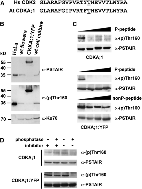
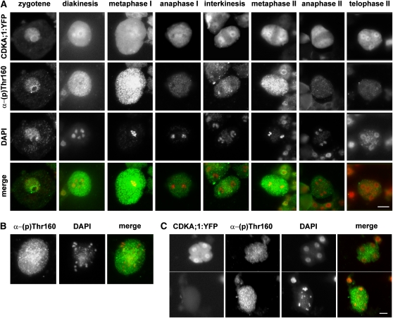
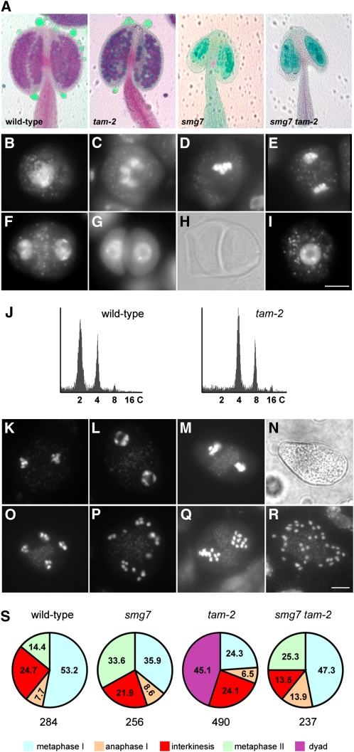
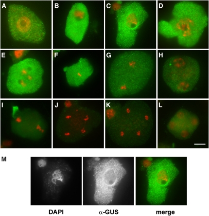
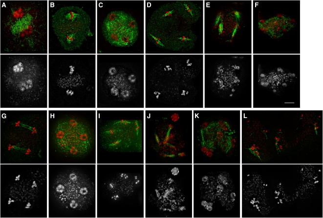
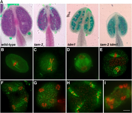
Meiosis is a modified cell division that produces four haploid nuclei from a single diploid cell in two rounds of chromosome segregation. Here, we analyze the role of Arabidopsis thaliana SUPPRESSOR WITH MORPHOGENETIC EFFECTS ON GENITALIA7 (SMG7), THREE DIVISION MUTANT1 (TDM1), and TARDY ASYNCHRONOUS MEIOSIS (TAM) in meiotic progression. SMG7 is a conserved nonsense-mediated mRNA decay factor that is also, in Arabidopsis, essential for completion of meiosis. Examination of activating CYCLIN DEPENDENT KINASE A;1 phosophorylation at Thr-161 suggests that the meiotic arrest observed in smg7 mutants is likely caused by a failure to downregulate cyclin-dependent kinase (CDK) activity at the end of the second meiotic division. Genetic analysis indicates that SMG7 and TDM1 act in the same pathway to facilitate exit from meiosis. We further demonstrate that the cyclin TAM is specifically expressed in meiosis I and has both stimulatory and inhibitory effects on progression to meiosis II. TAM knockouts skip the second meiotic division producing unreduced gametes, but inactivation of SMG7 or TDM1 alleviates TAM's requirement for entry into meiosis II. We propose a model that meiotic progression in Arabidopsis pollen mother cells is driven by a yet to be identified cyclin-CDK activity that is modulated by regulatory interactions between TDM1, SMG7, and TAM.


Figures

Figure 1.
Figure 1.
Immunoblot Analysis of CDKA;1 Phosphorylation at Thr-161. (A) Homology of Arabidopsis CDKA;1 and human CDK2 in the T-loop region with the indicated Thr residues at positions 161 and 160, respectively. (B) Immunodetection of phosphorylated Hs CDK2, At CDKA;1, and At CDKA;1:YFP proteins in extracts from HeLa cells, Arabidopsis wild-type influorescences and suspension culture, and CDKA;1:YFP flowers. (C) Immunodetection of Thr-161 phosphorylation of CDKA;1 and CDKA;1:YFP in the presence of increasing concentration (0, 1, 10, and 100 mg/mL) of phosphorylated (P) and nonphosphorylated (nonP) competitor peptides. Total CDKA;1 was detected with α-PSTAIR antibody as a loading control. (D) The Thr-161 phosphorylation signal is sensitive to phosphatase treatment. Protein extracts from wild-type and CDKA;1:YFP plants were pretreated with the lambda protein phosphatase in the presence or absence of phosphatase inhibitors as indicated. Proteins were analyzed by immunoblot with α-phospho-CDK2(Thr160) and α-PSTAIR antibody.
Figure 2.
Figure 2.
Immunolocalization of Thr-161 Phosphorylated CDKA;1 in the Course of Meiosis. (A) Meiocytes from PMCs of CDKA;1:YFP plants stained with α-phospho-CDK2(Thr160) antibody. DNA was counterstained with DAPI. Bar = 10 μm. (B) CDKA;1 Thr-161 phosphorylation in aberrant anaphase II of smg7 mutants. (C) Comparison of CDKA;1 Thr-161 phosphorylation signal in telophase II of CDKA;1:YFP plants (top panel) with aberrant anaphase II in smg7 mutants (bottom panel). PMCs from these plants were prepared on the same slide, and pictures of the meiocytes represent two cropped sections of the same photograph. CDKA;1:YFP meiocytes are distinguished from smg7 meiocytes by the presence of the YFP signal. Bars = 5 μM.
Figure 3.
Figure 3.
Epistasis Analysis of smg7 and tam-1 Mutations. (A) PMCs at interkinesis from wild-type and tam-1 mutants shown in phase contrast (top panel); nuclei are stained with DAPI (bottom panel). (B) Irregular behavior of meiotic chromosomes in PMCs of smg7 tam-1 double mutants. DNA is counterstained by DAPI. Bars in (A) and (B) = 5 μm. (C) Frequency (in %) of meiotic stages from metaphase I to metaphase II in wild-type, smg7-1, tam-1, and smg7 tam-1 plants. All plants were kept at 28°C during flowering. The total number of meiocytes is indicated below each pie chart.
Figure 4.
Figure 4.
SMG7 Deficiency Suppresses Premature Exit after Meiosis I Caused by the tam-2 Null Allele. (A) Pollen viability determined by Alexander staining of anthers. Viable pollen in wild-type and tam-2 plants is stained in red. No pollen is detected in anthers of smg7 and smg7 tam-2 mutants. (B) to (I) Meiotic chromosomes stained by DAPI in PMCs from tam-2 mutants. Zygotene (B), diakinesis (C), metaphase I (D), anaphase I (E), interkinesis (F), dyad visualized by DAPI staining (G) and by phase contrast (H), and microspore (I). Bar = 10 μm. (J) Flow cytometry analysis of DNA content in nuclei prepared from inflorescences of progeny of wild-type and tam-2 plants. (K) to (P) DAPI-stained PMCs from smg7 tam-2 double mutants. Anaphase I (K), interkinesis (L), fluorescence (M) and phase contrast (N) pictures of metaphase II, anaphase II (O), and irregular anaphase II (P). (Q) and (R) Metaphase II (Q) and irregular anaphase II (R) from tetraploid smg7 tam-2 double mutants. Bar = 5 μm (S) Frequency (in %) of meiotic stages from metaphase I to metaphase II in wild-type, smg7, tam-2, and smg7 tam-2 plants. The total number of meiocytes is indicated below each pie chart.
Figure 5.
Figure 5.
Immunolocalization of TAM:GUS in Meiosis. Fluorescein isothiocyanate signal detecting TAM:GUS is indicated in green and DNA counterstained with DAPI in red. Leptotene (A), zygotene (B), pachytene (C), diplotene (D), diakinesis (E), metaphase I (F), anaphase I (G), interkinesis (H), metaphase II (I), anaphase II ([J] and [K]), and telophase II (L). The pachytene in (M) is the same as in (C), shown as unmerged DAPI and fluorescein isothiocyanate pictures. Bar = 10 μm.
Figure 6.
Figure 6.
Meiosis in PMCs of tdm1 and smg7 tdm1 Mutants. The spindle was detected by immunostaining with anti-α-tubulin antibody (green), and DNA was counterstained with DAPI (red). Only DAPI staining is shown in the bottom panel. (A) to (F) Meiosis in tdm1 mutants. Interkinesis (A), metaphase II (B), telophase II (C), metaphase III (D), telophase III (E), and polyads (F). Bar = 5 μm. (G) to (L) Meiosis in smg7 tdm1 mutants. Anaphase II (G), telophase II (H), metaphase/anaphase III (I), anaphase III (J), polyads (K), and condensed chromatids aligned along spindles (L).
Figure 7.
Figure 7.
TDM1 Deficiency Suppresses Premature Exit after Meiosis I Caused by tam-2 Null Allele. (A) Pollen viability determined by Alexander staining of anthers. Viable pollen in wild-type and tam-2 plants is stained in red. No viable pollen is detected in tdm1 and tam-2 tdm1 mutants. (B) to (I) Meiosis in tam-2 tdm1 mutants. Spindle is shown in green and DNA counterstained with DAPI in red. Metaphase I (B), anaphase I (C), interkinesis (D), metaphase II (E), anaphase II (F), telophase II (G), metaphase/anaphase III (H), and polyads (I). Bar = 5 μm.
Figure 8.
Figure 8.
Models of CDK Regulation in Male Meiosis in the Wild Type and in tam-2, tam-1, and smg7 tam-1 Mutants. The wild type (A) and tam-2 (B), tam-1 (C), and smg7 tam-1 (D) mutants. The red line illustrates activity of the hypothetical CDK-cyclin kinase(s) in the course of meiosis. Green line depicts activity of TAM as inferred from TAM:GUS localization data. Stages of meioses and their approximate durations are indicated on the x axis (P1, prophase I; M1, metaphase I; A1, anaphase I; I/P2, interkinesis/prophase II; M2, metaphase II; A2, anaphase II). CDK activities and durations of meiotic stages reflect only hypothetical values and are not based on any exact measurements.

References

    1. Alexander M.P. (1969). Differential staining of aborted and nonaborted pollen. Stain Technol. 44: 117–122 - PubMed
    1. Averbeck N., Sunder S., Sample N., Wise J.A., Leatherwood J. (2005). Negative control contributes to an extensive program of meiotic splicing in fission yeast. Mol. Cell 18: 491–498 - PubMed
    1. Azumi Y., Liu D., Zhao D., Li W., Wang G., Hu Y., Ma H. (2002). Homolog interaction during meiotic prophase I in Arabidopsis requires the SOLO DANCERS gene encoding a novel cyclin-like protein. EMBO J. 21: 3081–3095 - PMC - PubMed
    1. Boruc J., Mylle E., Duda M., De Clercq R., Rombauts S., Geelen D., Hilson P., Inzé D., Van Damme D., Russinova E. (2010). Systematic localization of the Arabidopsis core cell cycle proteins reveals novel cell division complexes. Plant Physiol. 152: 553–565 - PMC - PubMed
    1. Carlile T.M., Amon A. (2008). Meiosis I is established through division-specific translational control of a cyclin. Cell 133: 280–291 - PMC - PubMed

Publication types

MeSH terms