. 2011 Mar 31;30(13):1566-76.
doi: 10.1038/onc.2010.532. Epub 2010 Nov 29.

Ligand-independent activation of c-Met by fibronectin and α(5)β(1)-integrin regulates ovarian cancer invasion and metastasis

Affiliations

Ligand-independent activation of c-Met by fibronectin and α(5)β(1)-integrin regulates ovarian cancer invasion and metastasis

A K Mitra et al. Oncogene. .

Abstract

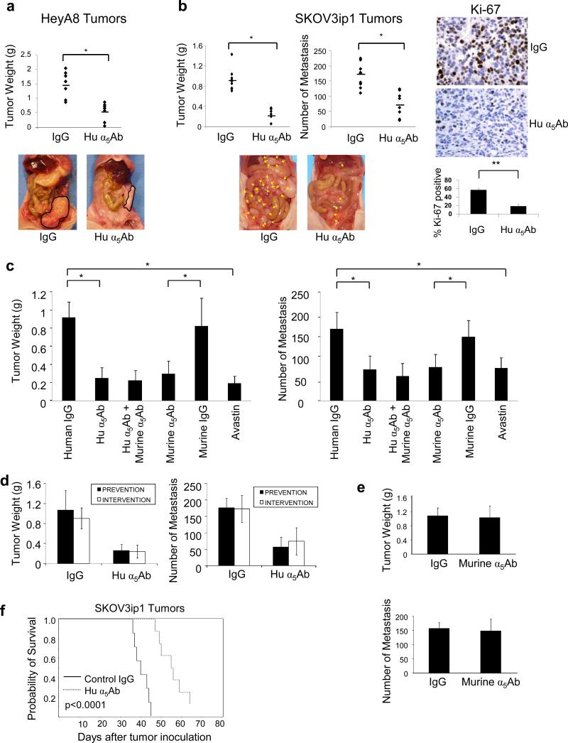
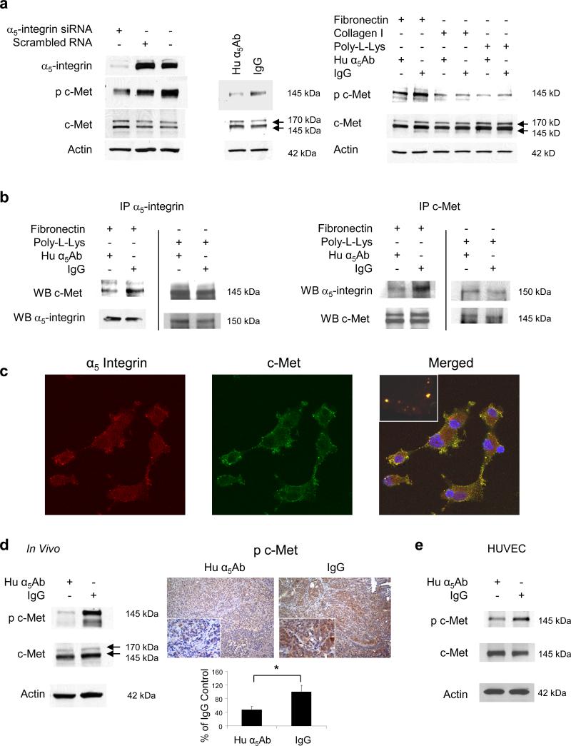
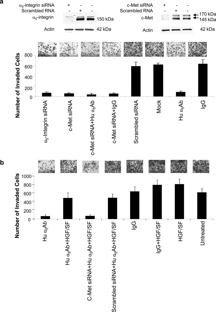
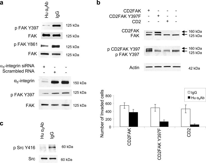
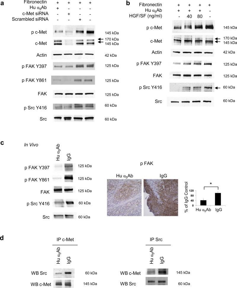
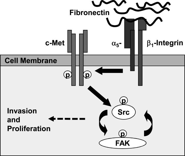
The role of the fibronectin receptor, α(5)β(1)-integrin, as an adhesion receptor and in angiogenesis is well established. However, its role in cancer cell invasion and metastasis is less clear. We describe a novel mechanism by which fibronectin regulates ovarian cancer cell signaling and promotes metastasis. Fibronectin binding to α(5)β(1)-integrin led to a direct association of α(5)-integrin with the receptor tyrosine kinase, c-Met, activating it in a hepatocyte growth factor/scatter factor (HGF/SF) independent manner. Subsequently, c-Met associated with Src, and activated Src and focal adhesion kinase (FAK). Inhibition of α(5)β(1)-integrin decreased the phosphorylation of c-Met, FAK and Src, both in vitro and in vivo. Independent activation of c-Met by its native ligand, HGF/SF, or overexpression of a constitutively active FAK in HeyA8 cells could overcome the effect of α(5)β(1)-integrin inhibition on tumor cell invasion, indicating that α(5)β(1)-integrin is upstream of c-Met, Src and FAK. Inhibition of α(5)β(1)-integrin on cancer cells in two xenograft models of ovarian cancer metastasis resulted in a significant decrease of tumor burden, which was independent of the effect of α(5)β(1)-integrin on angiogenesis. These data suggest that fibronectin promotes ovarian cancer invasion and metastasis through an α(5)β(1)-integrin/c-Met/FAK/Src-dependent signaling pathway, transducing signals through c-Met in an HGF/SF-independent manner.


Figures

Figure 1
Figure 1. Blocking α5β1-integrin in ovarian cancer cells inhibits metastasis in xenograft models
(a) HeyA8 cells (1×10⁶) were injected intraperitoneally (i.p.) in nude mice which were randomized into 2 groups of 10/group receiving 10mg/kg human α5β1-integrin antibody (Hu α5Ab) or control IgG. The effect of Hu α5Ab on tumor weight was assessed on day 20. (b) SKOV3ip1 cells (1×10⁶) were similarly injected i.p. treatment started on day 8 and tumor weight and the number of metastases assessed on day 35. Representative images of SKOV3ip1 tumors stained for Ki-67 (x400). Images of sections from 5 mice were quantified (**p<0.01). (c) SKOV3ip1 cells (1×10⁶) were injected i.p. in nude mice randomized into 6 groups of 10 each and were treated with the indicated antibodies starting on day 8. Mice were euthanized on day 35 and the effect on tumor weight and number of metastases was assessed. (*p<0.001, **p<0.01,). (d) The effect of co-administering SKOV3ip1 cells with a single dose of Hu α5Ab injected i.p. in mice (prevention) was compared with treatment of SKOV3ip1 tumor bearing mice starting 8 days after injection of cancer cells (intervention). The effect of treatment on tumor weight and number of metastases was measured. (e) A single dose of murine α5Ab was co-injected with SKOV3ip1 cells i.p. in nude mice (prevention) and tumor weight and number of metastases was measured on day 35. (f) Kaplan-Meier curve for survival. SKOV3ip1 cells were injected i.p. and 10 mice/group were treated with the human α5Ab or control IgG twice a week starting from day 8 (p<0.0001; log-rank test).
Figure 2
Figure 2. α5β1-Integrin signals through the receptor tyrosine kinase c-Met
(a) Left: HeyA8 cells were transfected with α5-integrin siRNA or treated with human α5Ab and plated on fibronectin followed by western blots for α5-Integrin, p-c-MetY1230,1234,1235 and c-Met. Right: HeyA8 cells were plated on fibronectin, collagen or poly-L-Lysine and were treated with human α5Ab followed by immunoblotting for phospho-c-Met Y1230,1234,1235, c-Met and actin. (b) HeyA8 cells were plated on fibronectin or poly-L-Lysine and treated with human α5Ab for 24 h. Cell lysates were immunoprecipitated (IP) with an antibody against α5-integrin or c-Met, respectively followed by immunoblotting (WB) for α5-integrin or c-Met. (c) Confocal microscopy. HeyA8 cells were plated on fibronectin coated cover slips, detected with c-Met and α5-integrin antibodies followed by fluorescein or Alexa Fluor 594 labeled secondary antibodies respectively. (d) SKOV3ip1 xenografts were treated with Hu α5Ab or IgG as described in Figure 1a. Left: Western blots for p-c-Met Y1230,1234,1235 and c-Met using tumor lysates. Right: Image of tumor sections stained for p-c-Met Y1200,1234,1235 (x200 and x400). Images of sections from 5 mice were quantified using Image J with color deconvolution (*p<0.001). (e) Human umbilical vein endothelial cells (HUVEC) were plated on fibronectin and treated with human α5Ab followed by western blots for p-c-MetY1230,1234,1235 and c-Met.
Figure 3
Figure 3. Inhibiting α5β1-integrin, silencing c-Met or both blocks invasion which can be overcome by activation of c-Met by HGF
(a) HeyA8 cells were transfected with the indicated siRNAs and seeded on matrigel coated Boyden chambers, treated with human α5Ab and allowed to invade for 24 h. Silencing was confirmed by immunoblotting. (b) HeyA8 cells or HeyA8 cells transfected with c-Met siRNA were placed in matrigel coated Boyden chambers, treated with human α5Ab with or without HGF/SF or untreated control as indicated and allowed to invade for 24h. For (a) and (b) the average number of invaded cells/field was plotted against the treatment given. Results are from three independent experiments.
Figure 4
Figure 4. Adhesion of ovarian cancer cells to fibronectin activates FAK and Src through α5β1-integrin
(a) Top: HeyA8 cells were transfected with α5-integrin siRNA or treated with human α5Ab and plated on fibronectin for 24h. Western blots were performed for p-FAK-Y397, p-FAK-Y861, FAK, and α5-integrin. (b) HeyA8 cells were transfected with CD2FAK (constitutively active FAK), CD2FAK-Y397F (mutated/inactive FAK) or CD2 (vector). Top: Expression and phosphorylation (Y397) of CD2FAK (160kDa) was verified by western blotting. Bottom: Transfected HeyA8 cells were added to matrigel coated Boyden chamber with human α5Ab or IgG and allowed to invade for 24 h. The number of invaded cells was counted. (c) HeyA8 cells were plated on fibronectin and treated with human α5Ab or IgG for 24 h. Phosphorylated (p)-Src Y416 and Src was detected by immunoblotting.
Figure 5
Figure 5. α5β1-Integrin signals through c-Met to activate FAK and Src
(a) HeyA8 cells were transfected with c-Met siRNA or with scrambled siRNA, plated on fibronectin and treated with human α5Ab. Cells were lysed, resolved on SDS-PAGE, transferred to nitrocellulose membrane and probed with the indicated antibodies. (b) HeyA8 cells were plated on fibronectin and treated with human α5Ab as indicated for 24 h. The cells were then treated with HGF/SF for 30 min and immunoblotting performed with the specified antibodies. Results in (a) and (b) are representative of three independent experiments. (c) SKOV3ip1 xenografts were treated with human α5Ab or with IgG as described in Figure 1 (b) Left: Tumor lysates were immunoblotted with the indicated antibodies. Right: Representative image of tumor sections stained for p-FAK-Y397 (x200). Images of sections from 5 mice were quantified using Image J (*p<0.001). (d) Immunoprecipitation. HeyA8 cells were plated on fibronectin and treated with human α5Ab or control IgG for 24 h followed by immunoprecipitation with an antibody against c-Met or Src, respectively followed by immunoblotting (WB) Src and c-Met. All results are representative of at least three independent experiments.
Figure 6
Figure 6. Fibronectin / α5β1-Integrin mediated regulation of c-Met
Proposed role of ligand independent activation of c-Met involving a fibronectin/α5β1-integrin/c-Met/Src/FAK dependent signaling pathway.

Comment in

References

    1. Avraamides C, Garmy-Susini B, Varner JA. Integrins in angiogenesis and lymphangiogenesis. Nature Rev Cancer. 2008:604–617. - PMC - PubMed
    1. Bhaskar V, Zhang D, Fox M, Seto P, Wong M, Wales PE, et al. A function blocking anti-mouse integrin α5β1 antibody inhibits angiogenesis and impedes tumor growth in vivo. J Transl Med. 2007;5:1–11. - PMC - PubMed
    1. Birchmeier C, Birchmeier W, Gherardi E, Vande Woude GF. Met, metastasis, motility and more. Nat Rev Mol Cell Biol. 2003;4:915–925. - PubMed
    1. Brader KR, Wolf JK, Hung M-C, Yu D, Crispens MA, van Golen KL, et al. Adenovirus E1A expression enhances the sensitivity of an ovarian cancer cell line to multiple cytotoxic agents through an apoptotic mechanism. Clin Cancer Res. 1997;3:2017–2024. - PubMed
    1. Caswell PT, Spence HJ, Parsons M, White DP, Clark K, Cheng KW, et al. Rab25 associates with α5β1 integrin to promote invasive migration in 3D microenvironments. Developmental Cell. 2007;13:496–510. - PubMed

Publication types