. 2011 Feb;19(2):362-71.
doi: 10.1038/mt.2010.263. Epub 2010 Nov 30.

MITF-siRNA formulation is a safe and effective therapy for human melasma

Affiliations

MITF-siRNA formulation is a safe and effective therapy for human melasma

Xiang Yi et al. Mol Ther. 2011 Feb.

Abstract

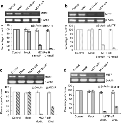
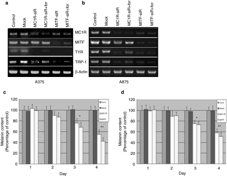
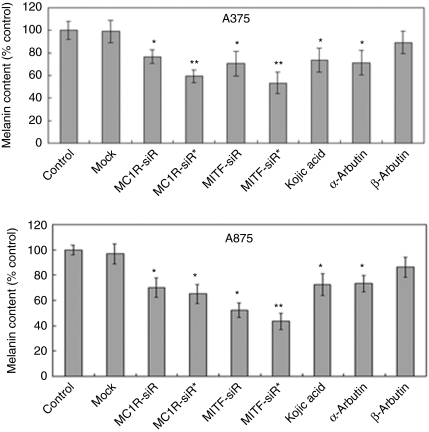
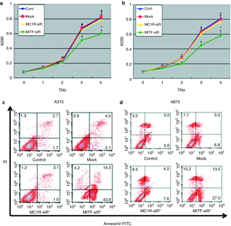
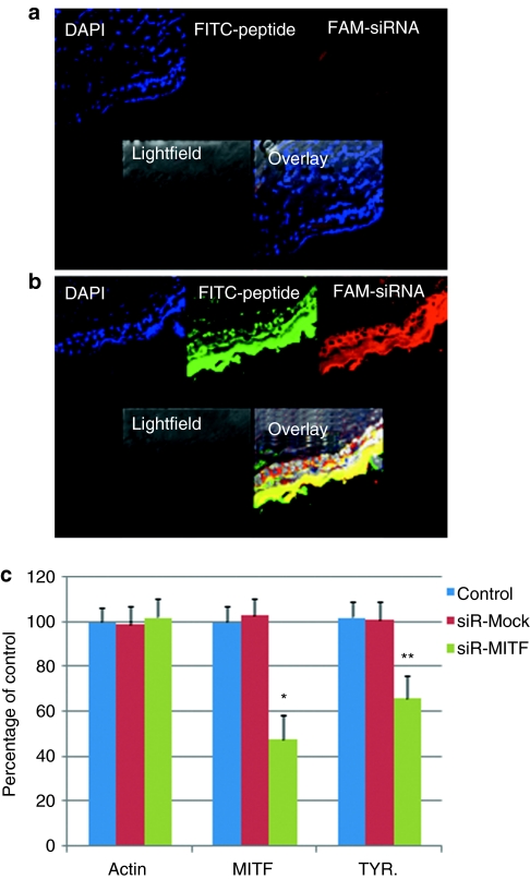
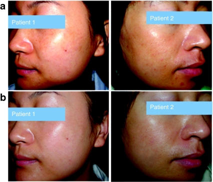
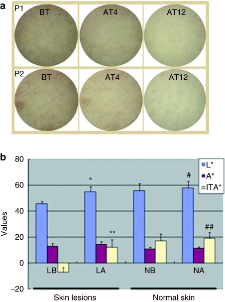
It is unclear whether siRNA-based agents can be a safe and effective therapy for diseases. In this study, we demonstrate that microphthalmia-associated transcription factor-siRNA (MITF-siR)-silenced MITF gene expression effectively induced a significant reduction in tyrosinase (TYR), tyrosinase-related protein 1, and melanocortin 1 receptor (MC1R) levels. The siRNAs caused obvious inhibition of melanin synthesis and melanoma cell apoptosis. Using a novel type of transdermal peptide, we developed the formulation of an MITF-siR cream. Results demonstrated that hyperpigmented facial lesions of siRNA-treated subjects were significantly lighter after 12 weeks of therapy than before treatment (P < 0.001); overall improvement was first noted after 4 weeks of siRNA treatment. At the end of treatment, clinical and colorimetric evaluations demonstrated a 90.4% lightening of the siRNA-treated lesions toward normal skin color. The relative melanin contents in the lesions and adjacent normal skin were decreased by 26% and 7.4%, respectively, after treatment with the MITF-siR formulation. Topical application of siRNA formulation significantly lightens brown facial hypermelanosis and lightens normal skin in Asian individuals. This treatment represents a safe and effective therapy for melasma, suggesting that siRNA-based agents could be developed for treating other diseases such as melanoma.


Figures

Figure 1
Figure 1
Melanocortin 1 receptor siRNA (MC1R-siR) and microphthalmia-associated transcription factor-siRNA (MITF-siR) significantly inhibit expression of their target genes. (a) When grown to 70% confluence in 6-well plates, melanoma cells were transfected with mock siRNA or MC1R-siR at concentrations of 5 nmol/l or 10 nmol/l for 24 hours. Subsequently, the cells were subjected to Trizol treatment. Reverse transcriptase–PCR was performed as described in Materials and Methods. β-Actin levels were a control for RNA loading. Relative levels of the expressed mc1r and β-actin mRNAs under various conditions were determined and normalized to their levels in the buffer control. Data are representative experiments performed in triplicate and are displayed as mean and SD. (b) Reverse transcriptase–PCR and quantitative analysis were employed for the MITF-siR case. (c) Reverse transcriptase–PCR to detect mc1r mRNA levels was performed on total RNAs from untreated melanoma cells (Control) or treated for 24 hours with mock siRNA or chemically modified MC1R-siR* plus TD1-R8 peptide or cholesterol conjugated MC1R-siR+ (Chol) alone. (d) The same protocol was used for the chemically modified MITF-siR* case.
Figure 2
Figure 2
Microphthalmia-associated transcription factor-siRNA (MITF-siR) significantly inhibits the expression of melanogenic genes and the synthesis of melanin in A375 and A875 melanoma cells. When (a) A375 and (b) A875 melanoma cells were at 70–80% confluence, they were transfected with 10 nmol/l mock siRNA or melanocortin 1 receptor siRNA (MC1R-siR) or MITF-siR for 24 hour, then treated without or with forskolin (for) at 40 µmol/l for a further 6 hours. Reverse transcriptase–PCR was performed as described in the Materials and Methods section. β-Actin levels were used as controls for RNA gel loading. A375 (c) and A875 (d) cells were treated once without or with the different siRNAs at 10 nmol/l. The melanin content was measured at different time points. Bars represent mean values ± SD of three independent experiments, each using duplicate culture flasks. *Significantly different from the nontreated control group at P < 0.01. **Significantly different from the untreated control group at P < 0.001.
Figure 3
Figure 3
Depigmenting agents inhibit the synthesis of melanin in A375 and A875 melanoma cells. Nine pools of melanoma cells were treated once with different agents such as different siRNAs at 10 nmol/l or other agents at 10 mmol/l, respectively. Three days after transfection, the cells were analyzed to determine the melanin content. Each tube contained 1 × 10⁶ cells. The melanin content of melanoma cells treated with buffer was the control. Bars represent mean values ± SD of three independent experiments, each using duplicate culture flasks. *Significantly different from the nontreated control group at P < 0.05. **Significantly different from the untreated control group at P < 0.001. MC1R-siR, melanocortin 1 receptor siRNA; MC1R-siR*, chemically modified melanocortin 1 receptor siRNA; MITF-siR, microphthalmia-associated transcription factor-siRNA; MITF-siR*, chemically modified microphthalmia-associated transcription factor-siRNA.
Figure 4
Figure 4
Microphthalmia-associated transcription factor-siRNA (MITF-siR) inhibits cell viability and induces apoptosis of melanoma cells. (a) A375 and (b) A875 melanoma cells were treated once per day for 3 days without or with different siRNAs at 10 mmol/l. Viabilities (a and b) and apoptosis (c and d) of the treated melanoma cells were determined using MTT assay and fluorescence-activated cell sorting analysis. Each point represents the mean ± SD Significant differences in cell viability between controls and agent-treated groups are indicated by #P < 0.01 or **P < 0.001. FITC, fluorescein isothiocyanate; MC1R-siR, melanocortin 1 receptor siRNA.
Figure 5
Figure 5
The transdermal peptide strongly facilitates the entry of microphthalmia-associated transcription factor-siRNA (MITF-siR) into the epidermis and dermis of mice. Fluorescence photomicrographs after topical application of MITF-siR cream formulation to hair-clipped BALB/c mouse skin after 2 hours. Pictures taken at ×40 magnification under a laser scanning confocal microscope. (a) A group of pictures for the group treated with naked siRNAs; (b) a group of pictures for the group treated with peptide-coated siRNA. Three animals were evaluated for each group; the micrographs show a representative section from one animal. Bar = 20 µm. (c) MITF-siR cream effectively inhibits MITF expression in vivo. Real-time PCR was performed as described in the Materials and Methods section. β-Actin levels were used as controls for RNA loading. Bars represent mean values ± SD of three independent experiments, each using duplicate mouse skin sized 1.5 × 0.2 cm2. * and **, significantly different from those in the mock group at P < 0.01 and P < 0.05, respectively. DAPI, 4′,6-diamidino-2-phenylindole; FITC, fluorescein isothiocyanate; TYR, tyrosinase.
Figure 6
Figure 6
Microphthalmia-associated transcription factor-siRNA (MITF-siR) cream significantly improves the images from two patients with facial melasma. (a) Before treatment. (b) After 12 weeks of treatment with MITF-siR cream, with lightening of pigmentation.
Figure 7
Figure 7
Microphthalmia-associated transcription factor-siRNA (MITF-siR) cream effectively inhibits melanin synthesis in vivo. (a) Dermoscopic pictures of melasma lesion before treatment (BT), and after 4 weeks (AT4) or 12 weeks (AT12) of treatment, showing complete improvement of skin lesions accompanied by lightening of the skin. P1 and P2 indicate patients 1 and 2, respectively. (b) Lightening (L*), red-green chromaticity coordinate (a*) and individual typology angle (ITA°) values for skin color changes before and after treatment. LB and LA refer to skin lesions before and after 12 weeks of treatment with MITF-siR cream; NB and NA refer to normal skin before and after 12 weeks of treatment, respectively. * and **, significantly different from those before treatment at P < 0.001. # and ##, significantly different from those before treatment at P < 0.05.

References

    1. Cestari T, Arellano I, Hexsel D., and, Ortonne JP, Latin American Pigmentary Disorders Academy Melasma in Latin America: options for therapy and treatment algorithm. J Eur Acad Dermatol Venereol. 2009;23:760–772. - PubMed
    1. Gupta AK, Gover MD, Nouri K., and, Taylor S. The treatment of melasma: a review of clinical trials. J Am Acad Dermatol. 2006;55:1048–1065. - PubMed
    1. Parvez S, Kang M, Chung HS, Cho C, Hong MC, Shin MK, et al. Survey and mechanism of skin depigmenting and lightening agents. Phytother Res. 2006;20:921–934. - PubMed
    1. Rafal ES, Griffiths CE, Ditre CM, Finkel LJ, Hamilton TA, Ellis CN, et al. Topical tretinoin (retinoic acid) treatment for liver spots associated with photodamage. N Engl J Med. 1992;326:368–374. - PubMed
    1. Stern RS. Clinical practice. Treatment of photoaging. N Engl J Med. 2004;350:1526–1534. - PubMed

Publication types

MeSH terms

Substances