. 2011 Mar;19(3):584-93.
doi: 10.1038/mt.2010.269. Epub 2010 Nov 30.

Generation of HIV-1 resistant and functional macrophages from hematopoietic stem cell-derived induced pluripotent stem cells

Affiliations

Generation of HIV-1 resistant and functional macrophages from hematopoietic stem cell-derived induced pluripotent stem cells

Amal Kambal et al. Mol Ther. 2011 Mar.

Abstract

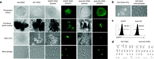
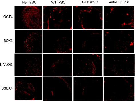
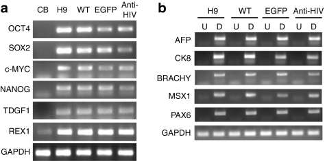
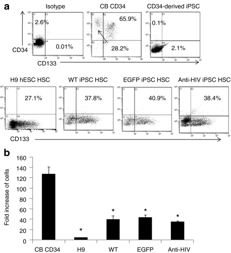
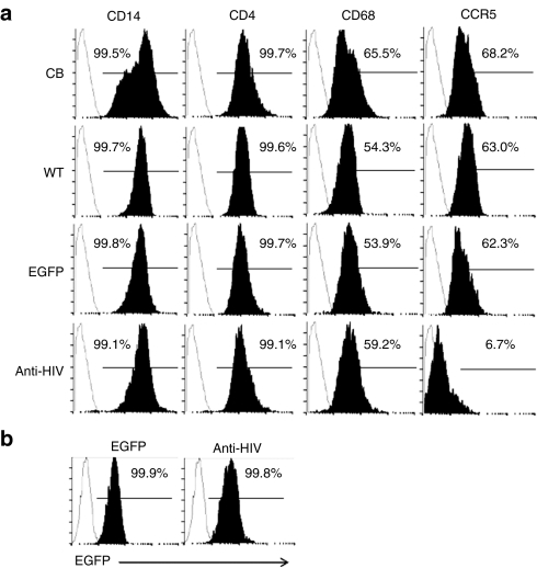
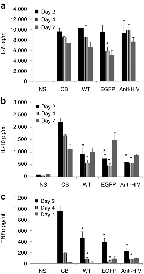
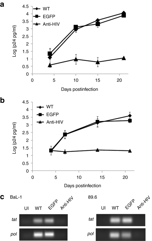
Induced pluripotent stem cells (iPSCs) have radically advanced the field of regenerative medicine by making possible the production of patient-specific pluripotent stem cells from adult individuals. By developing iPSCs to treat HIV, there is the potential for generating a continuous supply of therapeutic cells for transplantation into HIV-infected patients. In this study, we have used human hematopoietic stem cells (HSCs) to generate anti-HIV gene expressing iPSCs for HIV gene therapy. HSCs were dedifferentiated into continuously growing iPSC lines with four reprogramming factors and a combination anti-HIV lentiviral vector containing a CCR5 short hairpin RNA (shRNA) and a human/rhesus chimeric TRIM5α gene. Upon directed differentiation of the anti-HIV iPSCs toward the hematopoietic lineage, a robust quantity of colony-forming CD133(+) HSCs were obtained. These cells were further differentiated into functional end-stage macrophages which displayed a normal phenotypic profile. Upon viral challenge, the anti-HIV iPSC-derived macrophages exhibited strong protection from HIV-1 infection. Here, we demonstrate the ability of iPSCs to develop into HIV-1 resistant immune cells and highlight the potential use of iPSCs for HIV gene and cellular therapies.


Figures

Figure 1
Figure 1
Generation of anti-HIV induced pluripotent stem cells (iPSCs) and end-stage macrophages. (a) iPSCs were generated by transducing cord blood (CB) CD34+ human hematopoietic stem cells (HSCs) with four lentiviral vectors expressing the pluripotency factors octamer-binding transcription factor 4, sex determining region Y-box 2, Kruppel-like factor 4, and cytoplasmic Myc either alone [wild-type (WT)] or with an enhanced green fluorescent protein control vector (EGFP) or a combination anti-HIV vector (anti-HIV). iPSCs were further cocultured on OP9 stromal cells where cystic bodies developed. CD133+ HSCs were isolated from the cystic bodies and grown in semisolid methylcellulose media to form myeloid colony-forming units (CFUs). The CFUs were further cultured in media specific for macrophage development. EGFP and anti-HIV iPSCs and their differentiated progeny were visualized by both phase and EGFP fluorescence. H9 human embryonic stem cells (hESCs) and their differentiated progeny were used as controls. (b) CB CD34+ cells were used as a positive control and cultured in semisolid methylcellulose media and macrophage-specific media to derive myeloid CFUs and end-stage macrophages. (c) Representative fluorescence-activated cell sorting (FACS) plots displaying the EGFP percent of the EGFP and anti-HIV iPSCs at passage 21. WT iPSCs (EGFP negative) are displayed as the unshaded histogram. FACS analysis was performed in triplicate. (d) Representative karyotyping analyses of the WT and anti-HIV iPSCs derived from CB CD34+ HSCs. Karyotyping was performed in duplicate.
Figure 2
Figure 2
Expression of pluripotency markers by immunofluorescence. Induced pluripotent stem cells (iPSCs), wild-type (WT), enhanced green fluorescent protein (EGFP), and anti-HIV were stained with antibodies specific for the pluripotency markers octamer-binding transcription factor 4 (OCT4), sex determining region Y-box 2 (SOX2), NANOG, and stage-specific embryonic antigen-4 (SSEA4). Cells were visualized for fluorescence. H9 human embryonic stem cells (hESCs) were used as pluripotency positive controls. Pictures are representative of triplicate experiments.
Figure 3
Figure 3
Expression of pluripotency and differentiation genes by reverse transcriptase-PCR. (a) Total RNA from undifferentiated induced pluripotent stem cells (iPSCs) was extracted and analyzed by reverse transcriptase-PCR for the expression of the pluripotency genes octamer-binding transcription factor 4 (OCT4), sex determining region Y-box 2 (SOX2), cytoplasmic Myc (c-MYC), NANOG, teratocarcinoma-derived growth factor 1 (TDGF1), and REX1. CB CD34+ human hematopoietic stem cells (HSCs) were used as a control to detect expression in the “starter cells.” H9 human embryonic stem cells (hESCs) were used as a pluripotency positive control. (b) Total RNA was extracted from differentiated (D) cells from the cystic bodies which formed in the iPSC/OP9 cocultures and analyzed by RT-PCR for the expression of genes from all three germ layers including α-fetoprotein (AFP), cytokeratin 8 (CK8), brachyury (BRACHY), Msh homeobox 1 (MSX1), and paired box gene 6 (PAX6). Undifferentiated (U) cells were used as negative controls for expression. Undifferentiated and differentiated H9 hESCs were used as negative and positive controls, respectively. GAPDH was used as an internal loading control. Experiments were performed in duplicate. CB, cord blood; EGFP, enhanced green fluorescent protein; WT, wild type.
Figure 4
Figure 4
Detection and proliferation of induced pluripotent stem cells (iPSCs) derived CD133+ human hematopoietic stem cells (HSCs). (a) iPSCs, wild-type (WT), enhanced green fluorescent protein (EGFP), and anti-HIV were differentiated toward the hematopoietic lineage by OP9 cocultures. On day 9, the cells were analyzed for their expression of CD133 by FACS. Isotype control stained coculture cells are displayed in the top-left panel. CB CD34+ HSCs were used as a positive control for CD34 and CD133 expression (top-middle panel). Undifferentiated iPSCs were used as a negative control for CD34 and CD133 expression (top-right panel). Representative fluorescence-activated cell sorting plots are displayed from duplicate experiments. (b) Total cell counts were performed from methylcellulose cultured colony-forming units and analyzed for fold expansion of the input CD133+ cells. CB CD34+ HSCs were used as a positive comparative control. Asterisks above the bars indicate statistically significant values as compared to cord blood (CB) HSCs. Experiments were performed in triplicate. hESCs; human embryonic stem cells.
Figure 5
Figure 5
Phenotypic analysis of induced pluripotent stem cells (iPSCs) derived macrophages. (a) Macrophages derived from the wild-type (WT), enhanced green fluorescent protein (EGFP), and anti-HIV iPSCs were stained with antibodies specific for human CD14, CD4, CD68, and CCR5 and analyzed by fluorescence-activated cell sorting (FACS). Cord blood (CB) CD34+ HSC-derived macrophages were used as positive controls. Isotype controls are displayed as unshaded histograms. (b) EGFP and anti-HIV iPSC-derived macrophages were analyzed by FACS for EGFP expression. WT (EGFP) cells were used as negative controls and are displayed as unshaded histograms.
Figure 6
Figure 6
Cytokine secretion of induced pluripotent stem cells (iPSCs)-derived macrophages. Macrophages derived from the wild-type (WT), enhanced green fluorescent protein (EGFP), and anti-HIV iPSCs were stimulated with lipopolysaccharide. On days 2, 4, and 7, cell culture supernatants were analyzed for levels of secretion of (a) interleukin-6 (IL-6), (b) IL-10, and (c) tumor necrosis factor-α (TNFα). Cord blood (CB) CD34+ hematopoietic stem cells (HSC) derived macrophages were used as positive comparative controls. Asterisks above the bars indicate statistically significant values as compared to CB HSCs. Experiments were performed in triplicate. NS, nonstimulated.
Figure 7
Figure 7
HIV-1 challenge of induced pluripotent stem cells (iPSCs) derived macrophages. Macrophages derived from the wild type (WT) (diamonds), enhanced green fluorescent protein (EGFP) (squares), and anti-HIV (triangles) iPSCs were challenged with (a) an R5-tropic BaL-1 or (b) a dual-tropic 89.6 strain of HIV-1 at an multiplicity of infection of 0.05. On various days postinfection, cell culture supernatants were sampled and analyzed for p24 antigen by enzyme-linked immunosorbent assay. Experiments were performed in triplicate. (c) On day 21 postinfection, total RNA from uninfected (UI) and HIV-1 challenged macrophages were analyzed by reverse transcriptase-PCR for the expression of spliced (tat) and unspliced (pol) HIV-1 transcripts.

References

    1. Marcelin AG, Ceccherini-Silberstein F, Perno CF., and, Calvez V. Resistance to novel drug classes. Curr Opin HIV AIDS. 2009;4:531–537. - PubMed
    1. Martinez-Picado J, DePasquale MP, Kartsonis N, Hanna GJ, Wong J, Finzi D, et al. Antiretroviral resistance during successful therapy of HIV type 1 infection. Proc Natl Acad Sci USA. 2000;97:10948–10953. - PMC - PubMed
    1. Winters MA, Baxter JD, Mayers DL, Wentworth DN, Hoover ML, Neaton JD, et al. Frequency of antiretroviral drug resistance mutations in HIV-1 strains from patients failing triple drug regimens. The Terry Beirn Community Programs for Clinical Research on AIDS. Antivir Ther (Lond) 2000;5:57–63. - PubMed
    1. Lafeuillade A, Poggi C, Hittinger G., and, Chadapaud S. Phenotypic and genotypic resistance to nucleoside reverse transcriptase inhibitors in HIV-1 clinical isolates. HIV Med. 2001;2:231–235. - PubMed
    1. Strayer DS, Akkina R, Bunnell BA, Dropulic B, Planelles V, Pomerantz RJ, et al. Current status of gene therapy strategies to treat HIV/AIDS. Mol Ther. 2005;11:823–842. - PubMed

Publication types

MeSH terms