. 2010 Jun;4(3-4):142-52.
doi: 10.2976/1.3338713. Epub 2010 Apr 16.

From genes to neural tube defects (NTDs): insights from multiscale computational modeling

From genes to neural tube defects (NTDs): insights from multiscale computational modeling

G Wayne Brodland et al. HFSP J. 2010 Jun.

Abstract

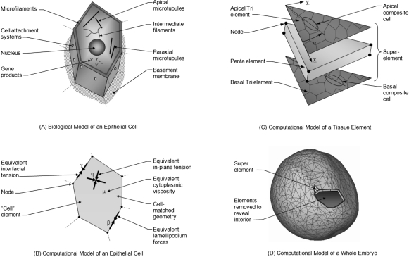
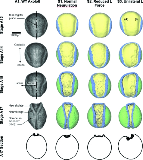
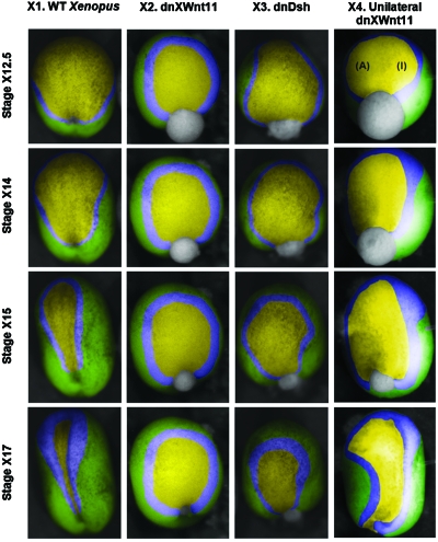
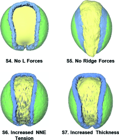
The morphogenetic movements, and the embryonic phenotypes they ultimately produce, are the consequence of a series of events that involve signaling pathways, cytoskeletal components, and cell- and tissue-level mechanical interactions. In order to better understand how these events work together in the context of amphibian neurulation, an existing multiscale computational model was augmented. Geometric data for this finite element-based mechanical model were obtained from 3D surface reconstructions of live axolotl embryos and serial sections of fixed specimens. Tissue mechanical properties were modeled using cell-based constitutive equations that include internal force generation and cell rearrangement, and equation parameters were adjusted manually to reflect biochemical changes including alterations in Shroom or the planar-cell-polarity pathway. The model indicates that neural tube defects can arise when convergent extension of the neural plate is reduced by as little as 20%, when it is eliminated on one side of the embryo, when neural ridge elevation is disrupted, when tension in the non-neural ectoderm is increased, or when the ectoderm thickness is increased. Where comparable conditions could be induced in Xenopus embryos, good agreement was found, an important step in model validation. The model reveals the neurulating embryo to be a finely tuned biomechanical system.


Figures

Figure 1
Figure 1. The multiscale computational model.
(A) Shown are primary components associated directly or indirectly with force generation. (B) In the corresponding cell-matched computational model, equivalent forces are calculated using equivalent joint loads and other engineering principles. Thousands of cell-level simulations involving tens to hundreds of cells such as the one shown were used to investigate the mechanics of embryonic epithelia. Ultimately, these studies provided sufficient understanding that cell-level constitutive equations relating stress, strain, cellular fabric, lamellipodium action and other relevant factors could be constructed. (C) These equations can be incorporated into “superelements” that can accurately represent the mechanics of a triangular piece of tissue. To model a whole embryo [Fig. 1D], its surface epithelium is broken into triangular regions consisting of several tens of cells. Each of these regions is represented by a superelement in which tri elements along the apical and basal surfaces of the monolayer tissue replicate the active forces produced by its cells. The angle α is measured as shown from an arbitrary reference edge. The penta element represents the passive forces generated by the cytoplasm and its contents (Chen and Brodland, 2008). (D) The initial geometry of the whole-embryo model was built by extruding triangles from a three-dimensional surface reconstruction of a live embryo, toward the centroid of the reconstruction a distance corresponding to the thickness of the ectoderm (surface layer) as seen in serial section sets. See text for details.
Figure 2
Figure 2. Comparison of model output with an axolotl embryo.
Column A1 shows photographs taken from a single live axolotl embryo, and a stage 17 cross-section from a different embryo. Stages in the figure are labeled with the letter A to denote them as axolotl stage numbers. Column S1 shows a simulation of normal neurulation. For the simulation shown in column S2, the strength of the lamellipodium action L was reduced to 80% of its normal value. Not only are the morphogenetic movements delayed but the tube does not close. When L forces are turned off on one side of the embryo (the side labeled with an I, for inactive, as opposed to the active side A), an asymmetric phenotype results (column S3), and closure fails. See Supplementary Material, for corresponding videos.
Figure 3
Figure 3. Experiments with Xenopus embryos.
Stage numbers are denoted with an X to distinguish them from the axolotl stage numbers shown in Fig. 2. Embryos injected with dnXWnt11 (column X2) or dnDsh constructs (column X3) or injected unilaterally with dnXWnt11 (column X4) are compared to controls (column X1) of the same clutch at developmental stages 12.5 (early neurulation), 14, 15, and 17 (late neurulation). Injected embryos show developmental defects including large and irregularly shaped neural plates, enlarged neural folds, and delayed neural fold migration. The manually overlaid colors correspond to those in Figs. 24.
Figure 4
Figure 4. Additional simulations.
When lamellipodium action is totally disabled in the model (Case S4), the resulting NTD embryo has features very similar to those produced when microinjected mRNA is used to disrupt the PCP pathway (Wallingford and Harland, 2002). When forces in the neural ridges assumed to be produced by Shroom are turned off in the model (Case S5), it predicts a final geometry consistent with that produced in Xenopus embryos when Shroom3 is disabled using an antisense morpholino oligonucleotide (Haigo et al., 2003). Case S6 shows an embryo in which the tensions in the NNE have been doubled and Case S7 shows an embryo in which the thickness of the epidermis and mesoderm have been doubled. All of the cases shown in this figure were run for extended periods of time and all give rise to NTDs as their final configurations, illustrated here, show.

References

    1. Belytschko, T, Liu, W K, and Moran, B (2000). Nonlinear Finite Elements for Continua and Structures, Wiley, New York.
    1. Bootsma, G J, and Brodland, G W (2005). “Automated 3-D reconstruction of the surface of live early-stage amphibian embryos.” IEEE Trans. Biomed. Eng. IEBEAX 52, 1407–1414.10.1109/TBME.2005.851500 - DOI - PubMed
    1. Bordzilovskaya, N P, Dettlaff, T A, Duhon, S T, and Malacinski, G M (1989). Developmental Biology of the Axolotl, Armstrong J B and Malacinski G M (eds), Oxford University Press, New York.
    1. Boyles, A L, Hammock, P, and Speer, M C (2005). “Candidate gene analysis in human neural tube defects.” Am. J. Med. Genet. C ZZZZZZ 135, 9–23.10.1002/ajmg.c.30048 - DOI - PubMed
    1. Brodland, G W (2002). “The differential interfacial tension hypothesis (DITH): a comprehensive theory for the self-rearrangement of embryonic cells and tissues.” ASME J. Biomech. Eng. JBENDY 124, 188–197.10.1115/1.1449491 - DOI - PubMed

LinkOut - more resources