. 2010 Nov 15:4:143.
doi: 10.3389/fnana.2010.00143. eCollection 2010.

Pulvinar projections to the striatum and amygdala in the tree shrew

Affiliations

Pulvinar projections to the striatum and amygdala in the tree shrew

Jonathan D Day-Brown et al. Front Neuroanat. .

Abstract

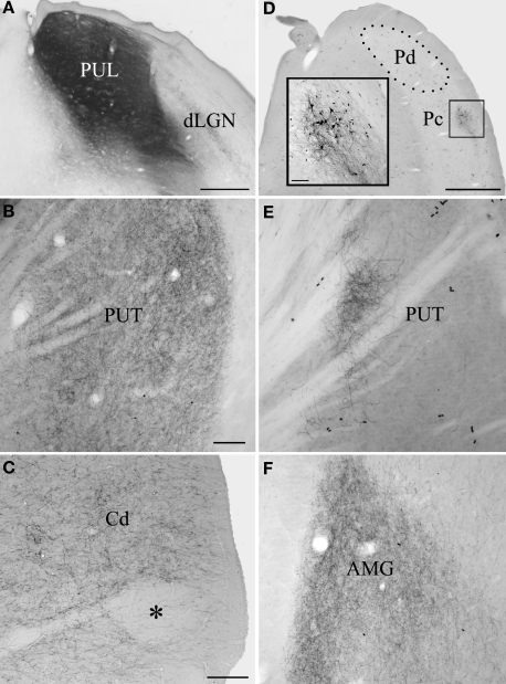
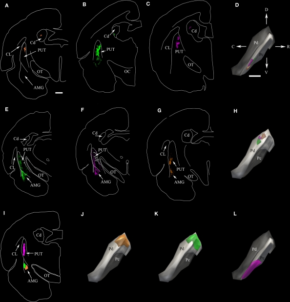
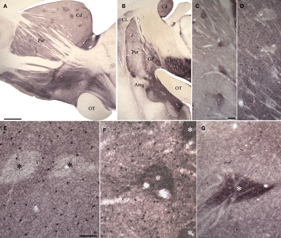
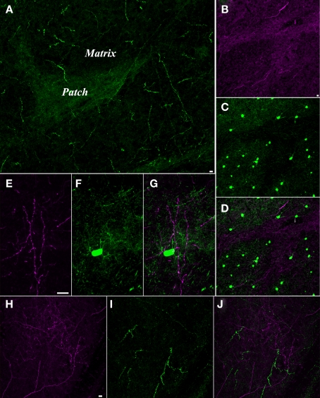
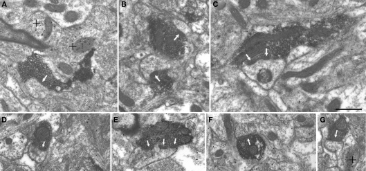
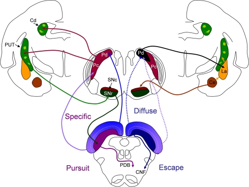
Visually guided movement is possible in the absence of conscious visual perception, a phenomenon referred to as "blindsight." Similarly, fearful images can elicit emotional responses in the absence of their conscious perception. Both capabilities are thought to be mediated by pathways from the retina through the superior colliculus (SC) and pulvinar nucleus. To define potential pathways that underlie behavioral responses to unperceived visual stimuli, we examined the projections from the pulvinar nucleus to the striatum and amygdala in the tree shrew (Tupaia belangeri), a species considered to be a prototypical primate. The tree shrew brain has a large pulvinar nucleus that contains two SC-recipient subdivisions; the dorsal (Pd) and central (Pc) pulvinar both receive topographic ("specific") projections from SC, and Pd receives an additional non-topographic ("diffuse") projection from SC (Chomsung et al., 2008). Anterograde and retrograde tract tracing revealed that both Pd and Pc project to the caudate and putamen, and Pd, but not Pc, additionally projects to the lateral amygdala. Using immunocytochemical staining for substance P (SP) and parvalbumin (PV) to reveal the patch/matrix organization of tree shrew striatum, we found that SP-rich/PV-poor patches interlock with a PV-rich/SP-poor matrix. Confocal microscopy revealed that tracer-labeled pulvino-striatal terminals preferentially innervate the matrix. Electron microscopy revealed that the postsynaptic targets of tracer-labeled pulvino-striatal and pulvino-amygdala terminals are spines, demonstrating that the pulvinar nucleus projects to the spiny output cells of the striatum matrix and the lateral amygdala, potentially relaying: (1) topographic visual information from SC to striatum to aid in guiding precise movements, and (2) non-topographic visual information from SC to the amygdala alerting the animal to potentially dangerous visual images.

Keywords: blindsight; matrix; striosome; superior colliculus; synapse.


Figures

Figure 1
Figure 1
The pulvinar nucleus projects to the striatum and lateral amygdala. Large BDA injections that involved both the dorsal (Pd) and central (Pc) pulvinar (A) labeled terminals throughout putamen (PUT, B) and caudate nucleus (Cd, C), although conspicuous patches (asterisk) were devoid of label. The injection illustrated in (A) also labeled terminals throughout the lateral amygdala (AMG, F). Smaller BDA injections confined to the Pd or Pc labeled restricted clusters of terminals in the Cd and PUT. An example Pc injection is illustrated in (D) (inset illustrates the injection site at higher magnification) and the resulting terminal distribution in the PUT in (E). Scale bar in A = 500 μm. Scale bar in B = 100 μm and also applies to (E). Scale bar in C = 100 μm and also applies to (F). Scale bar in D = 1 mm (inset scale bar = 100 μm). dLGN, dorsal lateral geniculate nucleus; OT, optic tract. A was previously published in Chomsung et al., .
Figure 2
Figure 2
The dorsal (Pd) and central (Pc) pulvinar project to the striatum; only the Pd projects to the amygdala. (A–C) Two dimensional (2D) neurolucida plots of the distribution of terminals labeled by anterograde transport in the caudate (Cd) and putamen (PUT) following three separate injections of BDA in the Pc. (D) Injection sites for (A–C) depicted in three-dimensional (3D) neurolucida reconstructions of the pulvinar nucleus. The color of each injection site corresponds to the color of the terminals in (A–C). (E–G) Two-dimensional plots of the distribution of terminals labeled by anterograde transport in the PUT and amygdala (AMG) following three separate injections of BDA in the Pd. (H) Three-dimension reconstructions of injection sites for (E–G). Because the full extent of the Pd is not viewable in the orientation depicted, the Pc subdivision was made transparent in this panel as well as in (J,K). (I) Two-dimension plots of the location of FG and CTB injections in the PUT and AMG. (J,K,L) Three-dimension reconstructions of the distribution of cells labeled in the pulvinar nucleus by retrograde transport from the injections illustrated in I. The color of the cell distributions corresponds to the injection site colors in I. Scale bar in A = 1 mm and applies to all 2D plots. Scale bar in D = 1 mm and applies to all 3D pulvinar reconstructions. C, caudal, CL, claustrum, D, dorsal, OC, optic chiasm, OT, optic tract, R, rostral, V, ventral. Orientation arrows in D also apply to all 3D pulvinar reconstructions.
Figure 3
Figure 3
Substance P (SP), parvalbumin (PV) and potassium channel interacting protein (KChIP) antibodies reveal patch/matrix organization of tree shrew striatum. (A,B) Sections (parasagittal, A; coronal, B) through the striatum stained for SP. Dark SP-positive patches are seen throughout the tree shrew striatum. (C,D) Adjacent sections through the putamen showing SP staining of patches (C) and PV staining in the matrix (D). (E) PV stains cells within the matrix but not within patches (black asterisks). (F) KChIP stains clusters of terminals in patches (white asterisk) and cells in the matrix. (G) SP stains densely distributed terminals in patches (white asterisk). In (A), scale bar = 1 mm and applies to B. Scale bar in C = 100 μm and applies to (D). Scale bar in E = 100 μm and applies to (F,G). Amg, amygdala, Cd, caudate, CL, claustrum, GP, globus pallidus, OT, optic tract, Put, putamen.
Figure 4
Figure 4
Pulvinar axons innervate the striatum matrix. (A) Confocal microscope image of substance P (SP, green) staining in the putamen. The matrix is sparsely innervated by thick SP axons, while the patches are densely innervated by fine SP axons. (B–D) Confocal image of a section stained for SP and parvalbumin (PV). The fine SP axons (purple, B) that innervate the patches interdigitate with a dense distribution of cells in the matrix that contain PV (C). (D) Merged image of (B) and (C) illustrates the interlocking arrangement of the SP-rich patches and the PV-rich matrix. (E–G) Pulvino-striatal axons innervate the PV-rich matrix. E illustrates BDA-labeled pulvino-striatal axons (purple) in the putamen. (F) shows a PV stained cell and dendrites (green) in the same section. (G) Merged image of (E) and (F) shows that pulvino-striatal axons target the PV-rich matrix. (H–J) Pulvino-striatal axons innervate the SP-poor matrix. (H) shows BDA-labeled pulvino-strital axons (purple) in the putamen. (I) shows SP staining (green) in the same section. Only the sparse SP axons of the matrix are stained. (J) A merged image of (H) and (I) illustrates that pulvino-striatal axons innervate the SP-poor matrix. All scale bars = 10 μm. Scale bar in (B) also applies to (C,D). Scale bar in (E) also applies to (F,G). Scale bar in (H) also applies to (I,J).
Figure 5
Figure 5
Pulvino-striatal and pulvino-amygdala terminals contact spines. Terminals labeled by the anterograde transport of BDA injected in the pulvinar nucleus contact (white arrows) spines in the putamen (A–C) and the amygdala (D–G). Single (A,B,D,F,G) perforated (C) and multiple (E) contacts were observed. Postembedding immunocytochemical staining for GABA revealed surrounding GABAergic terminals (high density of gold particles; + denotes) but BDA-labeled terminals and postsynaptic spines contained a low density of gold particles. Scale bar = 0.5 μm and applies to all panels.
Figure 6
Figure 6
The potential influence of dorsal (Pd) and central (Pc) pulvinar projections on basal ganglia circuits and behavior initiated by the superior colliculus (SC). Left side of figure: the Pd and Pc receive topographically arranged inputs from the SC (“specific”) and project to spiny output cells in the matrix (dark green) of the caudate (Cd) and putamen (PUT). GABAergic spiny cells of the Cd and PUT project to the substantia nigra pars reticulate (SNr) and GABAergic nigrotectal cells densely innervate the cells of origin of the crossed predorsal bundle (PDB). Tecto-pulvino-striatal projections could initiate pursuit movements mediated by the PDB. Right side of figure: the Pd receives additional non-topographic, convergent projections from the SC (“diffuse”) and projects to spiny output cells of the lateral amygdala (La). The La projects to the PUT and central amygdala (Ce). The Ce projects to the dopaminergic substantia nigra pars compacata (SNc). Tecto-pulvino-amygdala projections could serve to amplify escape movements mediated by the ipsilateral cuneiform (CNF) pathway. See text for details.

References

    1. Amorapanth P., LeDoux J. E., Nader K. (2000). Different lateral amygdala outputs mediate reactions and actions elicited by a fear-arousing stimulus. Nat. Neurosci. 3, 74–79 - PubMed
    1. Arend I., Machado L., Ward R., McGrath M., Ro T., Rafal R. D. (2008). The role of the human pulvinar in visual attention and action: evidence from temporal-order judgment, saccade decision, and antisaccade tasks. Prog. Brain Res. 171, 475–48310.1016/S0079-6123(08)00669-9 - DOI - PubMed
    1. Beckstead R. M. (1984). The thalamostriatal projection in the cat. J. Comp. Neurol. 223, 313–346 - PubMed
    1. Berman R. A., Wurtz R. H. (2008). Exploring the pulvinar path to visual cortex. Prog. Brain Res. 171, 467–47310.1016/S0079-6123(08)00668-7 - DOI - PMC - PubMed
    1. Berman R. A., Wurtz R. H. (2010). Functional identification of a pulvinar path from superior colliculus to cortical area MT. J. Neurosci. 30, 6342–635410.1523/JNEUROSCI.6176-09.2010 - DOI - PMC - PubMed

LinkOut - more resources