Tomographic assessment of the spine in children with spondylocostal dysotosis syndrome
- PMID: 21120293
- PMCID: PMC2972617
- DOI: 10.1590/s1807-59322010001000005
Tomographic assessment of the spine in children with spondylocostal dysotosis syndrome
Abstract
Objective: The aim of this study was to perform a detailed tomographic analysis of the skull base, craniocervical junction, and the entire spine in seven patients with spondylocostal dysostosis syndrome.
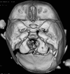
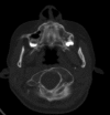
Method: Detailed scanning images have been organized in accordance with the most prominent clinical pathology. The reasons behind plagiocephaly, torticollis, short immobile neck, scoliosis and rigid back have been detected. Radiographic documentation was insufficient modality.
Results: Detailed computed tomography scans provided excellent delineation of the osseous abnormality pattern in our patients.
Conclusion: This article throws light on the most serious osseous manifestations of spondylocostal dysostosissyndrome.
Figures


Similar articles
-
Diffuse skull base/cervical fusion syndromes in two siblings with spondylocostal dysostosis syndrome: analysis via three dimensional computed tomography scanning.Spine (Phila Pa 1976). 2008 Jun 1;33(13):E425-8. doi: 10.1097/BRS.0b013e318175c2de. Spine (Phila Pa 1976). 2008. PMID: 18520929
-
Distinctive spine abnormalities in patients with Goldenhar syndrome: tomographic assessment.Eur Spine J. 2015 Mar;24(3):594-9. doi: 10.1007/s00586-014-3204-3. Epub 2014 Feb 7. Eur Spine J. 2015. PMID: 24504787
-
Prenatal sonographic features of spondylocostal dysostosis and diaphragmatic hernia in the first trimester.Ultrasound Obstet Gynecol. 1999 Mar;13(3):213-5. doi: 10.1046/j.1469-0705.1999.13030213.x. Ultrasound Obstet Gynecol. 1999. PMID: 10204217
-
Spondylocostal dysostosis: thirteen new cases treated by conservative and surgical means.Spine (Phila Pa 1976). 2004 Jul 1;29(13):1447-51. doi: 10.1097/01.brs.0000128761.72844.ab. Spine (Phila Pa 1976). 2004. PMID: 15223937 Review.
-
Three-dimensional ultrasound findings of spondylocostal dysostosis in the second trimester of pregnancy.Ultrasound Obstet Gynecol. 2006 May;27(5):580-2. doi: 10.1002/uog.2769. Ultrasound Obstet Gynecol. 2006. PMID: 16619382 Review.
References
-
- Bonafe L, Giunta C, Gassner M, Steinmann B, Superti Furga A. A cluster of autosomal recessive spondylocostal dysostosis caused by three newly identified DLL3 mutations segregating in a small village. Clin Genet. 2003;63:28–35. - PubMed
-
- Gellis SS, Feingold M. Spondylothoracic dysplasia (costovertebral dysplasia, Jarcho-Levin syndrome) Am J Dis Child. 1976;130:513–4. - PubMed
-
- Jarcho S, Levin PM. Hereditary malformations of the vertebral bodies. Johns Hopkins Med. 1938;62:216–26.
-
- Ramírez N, Cornier AS, Campbell RM, Carlo S, Arroyo S, Romeu J. Natural history of thoracic insufficiency syndrome: a spondylothoracic dysplasia perspective. J Bone Joint Surg Am. 2007;89:2663–75. - PubMed
-
- Maroteuax P, Le Merrer M. Medeicine-Sciences Flammarion; 2002. Maladies osseuses de l'enfant, 4th edn. Paris.
MeSH terms
LinkOut - more resources
Full Text Sources
Medical

