. 2011 Apr;39(4):1174-85.
doi: 10.1007/s10439-010-0217-x. Epub 2010 Dec 1.

Design of experiments approach to engineer cell-secreted matrices for directing osteogenic differentiation

Affiliations

Design of experiments approach to engineer cell-secreted matrices for directing osteogenic differentiation

Martin L Decaris et al. Ann Biomed Eng. 2011 Apr.

Abstract

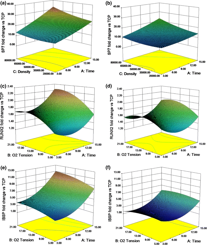
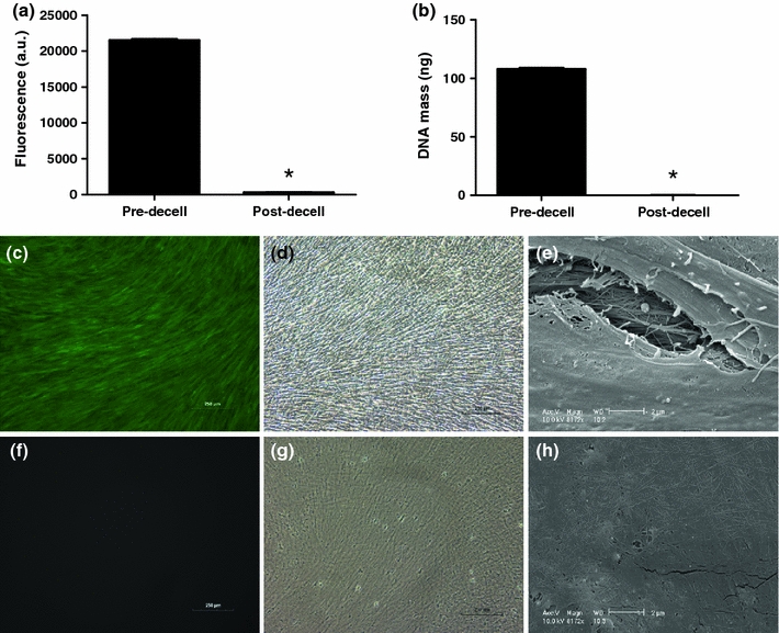
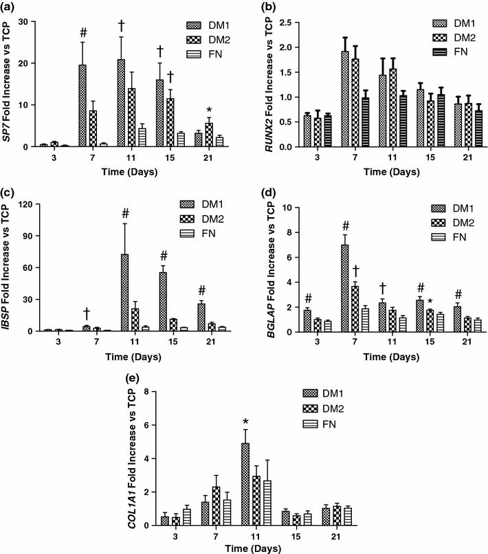
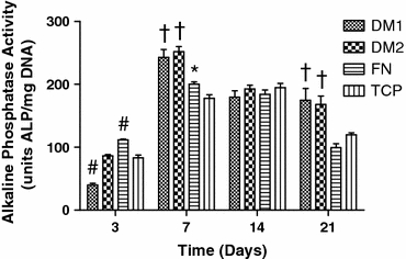
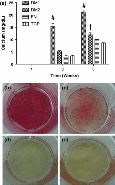
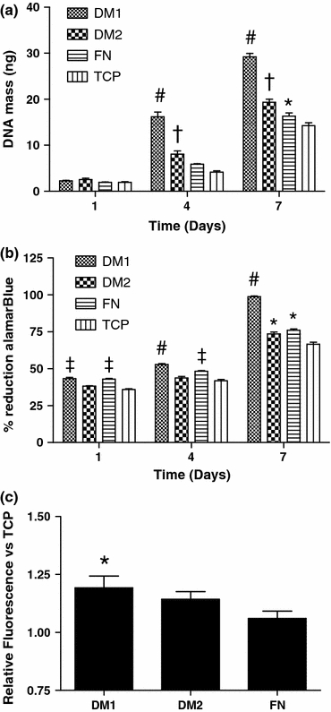
The presentation of extracellular matrix (ECM) proteins provides an opportunity to instruct the phenotype and behavior of responsive cells. Decellularized cell-secreted matrix coatings (DM) represent a biomimetic culture surface that retains the complexity of the natural ECM. Microenvironmental culture conditions alter the composition of these matrices and ultimately the ability of DMs to direct cell fate. We employed a design of experiments (DOE) multivariable analysis approach to determine the effects and interactions of four variables (culture duration, cell seeding density, oxygen tension, and media supplementation) on the capacity of DMs to direct the osteogenic differentiation of human mesenchymal stem cells (hMSCs). DOE analysis revealed that matrices created with extended culture duration, ascorbate-2-phosphate supplementation, and in ambient oxygen tension exhibited significant correlations with enhanced hMSC differentiation. We validated the DOE model results using DMs predicted to have superior (DM1) or lesser (DM2) osteogenic potential for naïve hMSCs. Compared to cells on DM2, hMSCs cultured on DM1 expressed 2-fold higher osterix levels and deposited 3-fold more calcium over 3 weeks. Cells on DM1 coatings also exhibited greater proliferation and viability compared to DM2-coated substrates. This study demonstrates that DOE-based analysis is a powerful tool for optimizing engineered systems by identifying significant variables that have the greatest contribution to the target output.


Figures

Figure 1
Figure 1
DMs engineered using extended culture duration, higher seeding densities, ambient oxygen tension, and A2P supplemented media appeared most effective at driving naïve hMSC osteogenesis, as determined by 3D surface model graphs exhibiting the correlation between culture conditions during matrix deposition and the resulting response of naïve hMSCs. (a) SP7 (osterix) expression in SM (21% O2), (b) SP7 expression in OM (21% O2), (c) RUNX2 expression in SM, (d) RUNX2 expression in OM, (e) IBSP expression in SM, and (f) IBSP expression in OM at 7 days. Data are fold change over expression in hMSCs on TCP
Figure 2
Figure 2
Quantitative analysis of the decellularization of DM1 utilizing (a) calcein uptake (n = 3) and (b) DNA quantification (n = 6). Fluorescent microscopy images of calcein uptake pre- (c) and post-decellularization (f). Bright field images (100× magnification) of cell layers pre- (d) and post-decellularization (g). SEM images of cell layers pre- (e) and post-decellularization (h) at 8172× magnification. Scale bars represent 250 μm (c, d, f, g) and 2 μm (e, h). *p < 0.0001 vs. pre-decell layers (a, b)
Figure 3
Figure 3
DM1 enhanced expression of several ostegenic marker genes in hMSCs compared to DM2 or control substrates when probed by qPCR in hMSCs cultured on each substrate for 3–21 days: (a) SP7, (b) IBSP, (c) RUNX2, (d) BGLAP (osteocalcin), and (e) COL1A1. Data were normalized to MRPL13 transcript level and reported as fold change in mRNA expression vs. TCP controls. # p < 0.05 vs. all groups, p < 0.05 vs. FN and TCP, *p < 0.05 vs. TCP only (n = 3)
Figure 4
Figure 4
Alkaline phosphatase activity from hMSCs cultured on each substrate for up to 21 days. # p < 0.05 vs. all groups, p < 0.05 vs. FN and TCP, *p < 0.05 vs. TCP only (n = 4–6)
Figure 5
Figure 5
hMSCs cultured on DM1 exhibited increased calcium deposition compared to cells on DM2 or control substrates. (a) Calcium deposited from hMSCs cultured on each substrate for 1, 3, and 5 weeks. Alizarin red staining of fixed hMSC layers cultured for 3 weeks on (b) DM1, (c) DM2, (d) FN, and (e) TCP. # p < 0.05 vs. all groups, p < 0.05 vs. FN and TCP (n = 3–4)
Figure 6
Figure 6
hMSC proliferation and viability are enhanced when cultured on DM1 compared to culture on DM2 or control substrates. (a) Total DNA on each substrate at 1, 4, and 7 days post-seeding. (b) AlamarBlue reduction by hMSCs cultured on each substrate at 1, 4, and 7 days. (c) Calcein uptake by hMSCs seeded on each substrate and under environmental stress for 24 h. # p < 0.05 vs. all groups, p < 0.05 vs. DM2 and TCP, p < 0.05 vs. FN and TCP, *p < 0.05 vs. TCP only (n = 4–6)
Figure 7
Figure 7
Calcein uptake by hMSCs seeded on each substrate at 1 and 4 h post-seeding. # p < 0.05 vs. all groups, p < 0.05 vs. FN and TCP, *p < 0.05 vs. TCP only (n = 4)

Similar articles

Cited by

References

    1. Aulin C, Foroughi F, Brown R, Hilborn J. Extracellular matrix-polymer hybrid materials produced in a pulsed-flow bioreactor system. J. Tissue Eng. Regen. Med. 2009;3:188–195. doi: 10.1002/term.152. - DOI - PubMed
    1. Badylak SF, Freytes DO, Gilbert TW. Extracellular matrix as a biological scaffold material: structure and function. Acta Biomater. 2009;5:1–13. doi: 10.1016/j.actbio.2008.09.013. - DOI - PubMed
    1. Badylak SF, Gilbert TW. Immune response to biologic scaffold materials. Semin. Immunol. 2008;20:109–116. - PMC - PubMed
    1. Bancroft GN, Sikavitsas VI, van den Dolder J, Sheffield TL, Ambrose CG, Jansen JA, Mikos AG. Fluid flow increases mineralized matrix deposition in 3D perfusion culture of marrow stromal osteoblasts in a dose-dependent manner. Proc. Natl. Acad. Sci. USA. 2002;99:12600–12605. doi: 10.1073/pnas.202296599. - DOI - PMC - PubMed
    1. Bennett KP, Bergeron C, Acar E, Klees RF, Vandenberg SL, Yener B, Plopper GE. Proteomics reveals multiple routes to the osteogenic phenotype in mesenchymal stem cells. BMC Genomics. 2007;8:380. doi: 10.1186/1471-2164-8-380. - DOI - PMC - PubMed

Publication types

MeSH terms