. 2011 Jun;32(6):579-89.
doi: 10.1002/humu.21406. Epub 2011 May 5.

Molecular defects in human carbamoy phosphate synthetase I: mutational spectrum, diagnostic and protein structure considerations

Affiliations

Molecular defects in human carbamoy phosphate synthetase I: mutational spectrum, diagnostic and protein structure considerations

Johannes Häberle et al. Hum Mutat. 2011 Jun.

Abstract

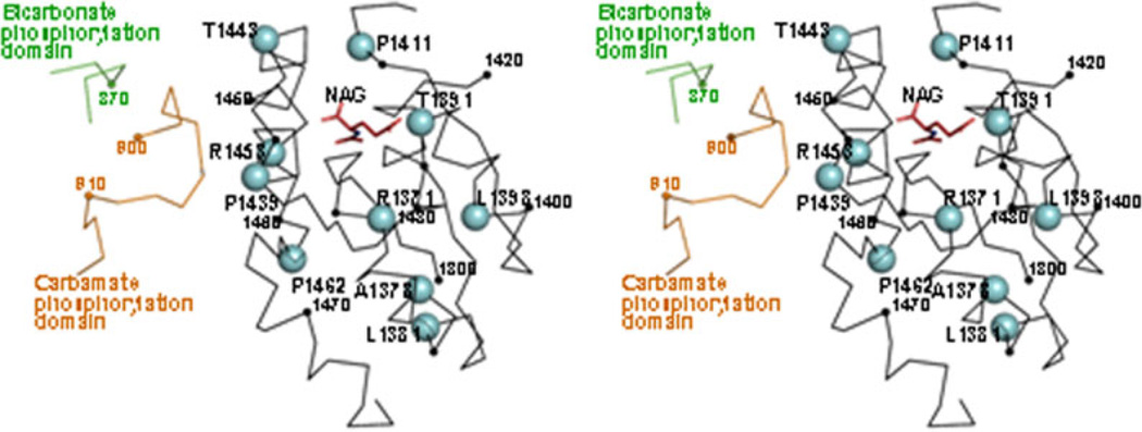
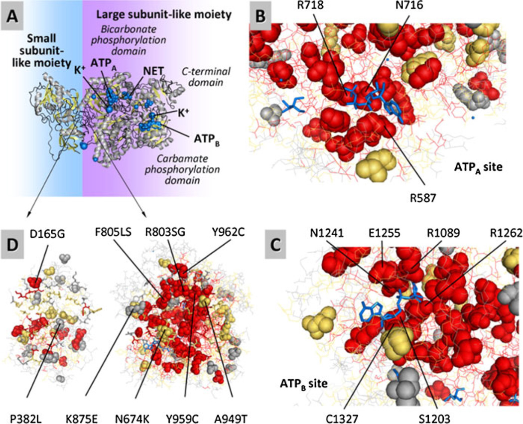
Deficiency of carbamoyl phosphate synthetase I (CPSI) results in hyperammonemia ranging from neonatally lethal to environmentally induced adult-onset disease. Over 24 years, analysis of tissue and DNA samples from 205 unrelated individuals diagnosed with CPSI deficiency (CPSID) detected 192 unique CPS1 gene changes, of which 130 are reported here for the first time. Pooled with the already reported mutations, they constitute a total of 222 changes, including 136 missense, 15 nonsense, 50 changes of other types resulting in enzyme truncation, and 21 other changes causing in-frame alterations. Only ∼10% of the mutations recur in unrelated families, predominantly affecting CpG dinucleotides, further complicating the diagnosis because of the "private" nature of such mutations. Missense changes are unevenly distributed along the gene, highlighting the existence of CPSI regions having greater functional importance than other regions. We exploit the crystal structure of the CPSI allosteric domain to rationalize the effects of mutations affecting it. Comparative modeling is used to create a structural model for the remainder of the enzyme. Missense changes are found to directly correlate, respectively, with the one-residue evolutionary importance and inversely correlate with solvent accessibility of the mutated residue. This is the first large-scale report of CPS1 mutations spanning a wide variety of molecular defects highlighting important regions in this protein.


Figures

Figure 1
Figure 1
Distribution of mutations among the 38 CPS1 exons and correspondence with the domains of the protein. The CPSI coding sequence is schematized as a horizontal bar at the middle, with segments therein corresponding to individual exons (numbered; odd exons are shadowed gray). A parallel bar of the same length at the bottom schematizes the enzyme polypeptide, giving in different color the various enzyme domains, which are shown in exact correspondence with the exons encoding them: LP, mitochondrial targeting peptide (not present in mature CPSI). ISD and GSD, the two subdomains of the 40-kDa CPSI region corresponding to the small subunit of E. coli CPS. In the latter enzyme the ISD and the GSD are, respectively, the intersubunit interaction domain and a glutaminase domain. The glutaminase activity is lost in human CPSI. BPSD, UFSD, CPSD, and ASD, the four domains of the 120-kDa region of CPSI corresponding to the E. coli CPS large subunit. BPSD and CPSD, respective bicarbonate and carbamate phosphorylation domains. ASD, allosteric domain (the domain that binds NAG). UFSD, unknown function domain. The steps of the CPSI reaction are schematized below the protein, placing the two phosphorylation domains below the residues catalyzing them. Planted on the exons of the coding sequence, the banners in blue localize the missense mutations summarized in Supp. Table S1. Inverted banners below the coding sequence give the other types of mutations, in the color code as indicated at the figure foot. Novel mutations are boldface and italicized. Mutations with known neonatal-onset CPSID are shown in red font.
Figure 2
Figure 2
Density of missense mutations per CPS1 exon or CPSI domain and correlation with the domain structure of the enzyme. A: Bar representation of the CPS1 coding sequence/CPSI protein to show the exon and domain composition and the different sizes of the various exons and domains, superimposing on them vertical lines corresponding to the exact locations of the different missense changes summarized in Table 1. The bar has the same length as the 4500-base coding sequence or 1500-residue amino acid sequence at the X-axes of the plots below. It is divided into the various exons, which are represented to scale and numbered. The different domains along the CPSI protein are superimposed on the exon bar, again in exact representation of sizes and boundaries (given as the number of the amino acid residue in the protein sequence), coloring each domain as in the bar at the bottom of Figure 1. The full domain name is shown above the bar in its appropriate color. Question marks indicate unknown function. The span in the polypeptide of the 40-kDa and 120-kDa moieties of the protein and of the two homologous repeats that make up the 120-kDa moiety are shown above the bar. The setting is such that there is exact physical correspondence between the exons and the domains in the bar and the two plots below it. B: The number of missense mutations (from Table 1) per exon, normalized per 100 nucleotides, is plotted along the coding sequence, with the symbols placed at the middle point of each exon (some representative exon numbers are given). Thin line, all missense mutations. Thick line and large filled triangles, missense mutations that do not fall on CpG dinucleotides. C: Bar plot of the number of missense mutations per domain or subdomain, normalized per 100 amino acids, along the amino acid sequence of CPSI. The width of each bar corresponds to the number of residues in the domain. The colors and names of the bars correspond to the naming and coloring of the domains that are given at the bottom of Figure 1. The shadowed bars that are superimposed on both phosphorylation domains, BPSD and CPSD, correspond to the frequency of mutations in the three subdomains A, B, and C, that compose each of these phosphorylation domains. Horizontal broken lines, number of missense mutations that do not fall on CpG dinucleotides.
Figure 3
Figure 3
Structure of the C-terminal domain of CPSI, with indication of the missense mutations found within this domain. Stereo view of the crystal structure of this domain (Cα trace in black) produced in a cell-free system (Protein DataBank file 2yvq), with fitting in the structure of the bound NAG molecule (in sticks and colored) [Pekkala et al., 2009]. The green and brown loops belong, respectively, to the bicarbonate and carbamate phosphorylation domains of the superimposed structure of E. coli CPS. Small spheres mark every tenth residue (numbering some of them, in small type). Large cyan spheres correspond to the indicated allosteric domain residues, which are those at which missense mutations have been found (Supp. Table S1). With the purpose of comparison, the projection and mode of representation are as similar as possible to those in Figure 1D of [Pekkala et al., 2010].
Figure 4
Figure 4
Structural model of human CPSI and its binding sites, predicting the localization of the mutated residues and providing an indication of the evolutionary importance (EI) of each residue. A: A ribbon representation of the enzyme, with ligands represented by blue spheres. The model was built from the structure of E. coli CPS using SWISS-MODEL (see Methods and Supporting Information). Similarly to the E. coli template structure, the BPSD and CPSD domains are shown binding AMPPNP, an inert ATP analogue labeled as ATPA and ATPB in the BPSD and CPSD subdomains, respectively. The NH3 analogue, a tetraethylammonium ion, is labeled as NET and is located in the BPSD subdomain. In the human CPSI ornithine is not considered to be an effector, but it is an activator of E. coli CPS. It has been placed here at the equivalent location in the model, between the CPSD and ASD subdomains. B–D: Detailed view of the CPSD and BPSD subdomains. Residues with high, medium, and low evolutionary importance are colored as red, orange, and gray, respectively. Ligands are shown blue, in sticks representation. The majority of the missense mutations affect evolutionary important residues and occur internally (low solvent accessibility). Sticks and lines represent nonmutated residues at and away from the binding site of interest, respectively. B: Bicarbonate phosphorylation subdomain binding ATPA. C: Carbamate phosphorylation subdomain binding ATPB. D: Missense mutation affecting amino acid residues predicted to play role in the interaction between the small subunit-like and large subunit-like moieties of CPSI.
Figure 5
Figure 5
The summary of all CPS1 mutations reported to date.

References

    1. Ahuja V, Powers-Lee SG. Human carbamoyl-phosphate synthetase: insight into N-acetylglutamate interaction and the functional effects of a common single nucleotide polymorphism. J Inherit Metab Dis. 2008;31:481–491. - PubMed
    1. Alonso E, Cervera J, Garcia-Espana A, Bendala E, Rubio V. Oxidative inactivation of carbamoyl phosphate synthetase (ammonia). Mechanism and sites of oxidation, degradation of the oxidized enzyme, and inactivation by glycerol, EDTA, and thiol protecting agents. J Biol Chem. 1992;267:4524–4532. - PubMed
    1. Alonso E, Rubio V. Affinity cleavage of carbamoyl-phosphate synthetase I localizes regions of the enzyme interacting with the molecule of ATP that phosphorylates carbamate. Eur J Biochem. 1995;229:377–384. - PubMed
    1. Aoshima T, Kajita M, Sekido Y, Kikuchi S, Yasuda I, Saheki T, Watanabe K, Shimokata K, Niwa T. Novel mutations (H337R and 238-362del) in the CPS1 gene cause carbamoyl phosphate synthetase I deficiency. Hum Hered. 2001a;52:99–101. - PubMed
    1. Aoshima T, Kajita M, Sekido Y, Mimura S, Itakura A, Yasuda I, Saheki T, Watanabe K, Shimokata K, Niwa T. Carbamoyl phosphate synthetase I deficiency: molecular genetic findings and prenatal diagnosis. Prenat Diagn. 2001b;21:634–637. - PubMed

Publication types

MeSH terms

Substances