. 2010 Dec 22;132(50):17961-72.
doi: 10.1021/ja1087612. Epub 2010 Dec 1.

Processive replication of single DNA molecules in a nanopore catalyzed by phi29 DNA polymerase

Affiliations

Processive replication of single DNA molecules in a nanopore catalyzed by phi29 DNA polymerase

Kate R Lieberman et al. J Am Chem Soc. .

Abstract

Coupling nucleic acid processing enzymes to nanoscale pores allows controlled movement of individual DNA or RNA strands that is reported as an ionic current/time series. Hundreds of individual enzyme complexes can be examined in single-file order at high bandwidth and spatial resolution. The bacteriophage phi29 DNA polymerase (phi29 DNAP) is an attractive candidate for this technology, due to its remarkable processivity and high affinity for DNA substrates. Here we show that phi29 DNAP-DNA complexes are stable when captured in an electric field across the α-hemolysin nanopore. DNA substrates were activated for replication at the nanopore orifice by exploiting the 3'-5' exonuclease activity of wild-type phi29 DNAP to excise a 3'-H terminal residue, yielding a primer strand 3'-OH. In the presence of deoxynucleoside triphosphates, DNA synthesis was initiated, allowing real-time detection of numerous sequential nucleotide additions that was limited only by DNA template length. Translocation of phi29 DNAP along DNA substrates was observed in real time at Ångstrom-scale precision as the template strand was drawn through the nanopore lumen during replication.


Figures

Figure 1
Figure 1
Capture of polymerase-DNA binary complexes in the α-HL nanopore. (a) Schematic of the nanopore device. A single α-HL nanopore is inserted in a 30 µm-diameter lipid bilayer that separates two 100 µL wells containing 10 mM K-Hepes, pH 8.0, 300 mM KCl, 1 mM DTT and 1 mM EDTA at 23 °C. The nanopore buffer contained no added MgCl2. A membrane potential across the bilayer is determined by AgCl electrodes in series with an Axon 200B amplifier. (b) DNA hairpin substrate used in this experiment. The DNA strand is designed to fold back onto itself forming a 14 bp duplex stem joined by a four dTMP residue loop. The 3′ residue of the primer strand is ddCMP. The red Xs indicate the five abasic (1′,2′-H) residues that span positions +12 to +16 of the DNA template strand (indicated by numbered arrows above the sequence). Template strand numbering is relative to the first unpaired residue (dCMP) residue at position n = 0 (indicated in blue). The chemical structure of an abasic monomer is shown below the DNA sequence. (c) Ionic current signature for capture of a KF(exo-)-DNA complex at 180 mV applied potential. (i) is the open channel current; (ii) is the enzyme bound state current (IEBS); (iii) is the current caused when voltage-promoted dissociation of KF(exo-) from the DNA causes the duplex segment of the hairpin to drop into the pore vestibule; (iv) is the return to open channel current caused by unzipping of the DNA hairpin while it is within the nanopore vestibule followed by electrophoresis to the trans compartment. Median EBS dwell time for the KF(exo-) binary complexes was 1.9 ms (n = 199), identical to the dwell time for binary complexes formed with the same hairpin substrate in the presence of 5 mM MgCl2. (d) Ionic current signature upon capture of a phi29 DNAP-DNA complex at 180 mV potential. (i) is the open channel current; (ii) is IEBS for the phi29 DNAP-DNA binary complex; (iii) is a terminal cascade of the current caused by putative unzipping of the DNA duplex while it is bound to phi29 DNAP, and the consequent ratcheting of the DNA through the pore; and (iv) is the restoration of the open channel current following electrophoresis of the unzipped DNA to the trans compartment. The concentrations of KF(exo-) in panel c and phi29 DNAP in panel d were 0.75 µM; in both panels the DNA concentration was 1.0 µM. Note the difference in time scale between panels c and d.
Figure 2
Figure 2
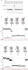
Duplex unzipping during DNA hairpin dissociation from phi29 DNAP at 180 mV applied potential is reversed at 70 mV. (a) Protection in the bulk phase of a 14 bp DNA hairpin substrate from phi29 DNAP-catalyzed 3′-5′ exonucleolytic degradation by a ddNMP (3′-H) terminated primer strand. Hairpin substrates (1 µM) labeled with 5′-6-FAM bearing either a 3′-OH (lanes 1–6) or 3′-H (lanes 7–12) terminus were incubated at room temperature with 0.75 µM phi29 DNAP in buffer containing 10 mM K-Hepes, pH 8.0, 300 mM KCl, 1 mM DTT and 1 mM EDTA for the times indicated. The reactions in lanes 1 and 7 contained no added MgCl2; those in lanes 2–6 and 8–12 contained 10 mM MgCl2. The reactions in lanes 6 and 12 also contained 200 µM each dATP, dCTP, dGTP and dTTP. Reaction products were resolved on an 18% denaturing polyacrylamide gel. Positions of the gel bands corresponding to the intact 67 mer starting substrates and the 102 mer full-length extension products are indicated with arrows on the side of the gel. Sequences of the 5′-6-FAM labeled DNA hairpins are shown in Figure S1. (b) Steps in the pathway of voltage-promoted phi29 DNAP-DNA complex dissociation are reversible. In this experiment, the buffer contained 1mM EDTA and no added MgCl2 in order to prevent phi29 DNAP 3′-5′ exonucleolytic activity. (i) Capture of a phi29 DNAP-DNA binary complex formed with the hairpin substrate shown in Figure 1b. This positions the abasic insert, located between positions +12 to +16 of the template strand, in the limiting aperture of the nanopore lumen, yielding an IEBS of 35 pA; (ii) after several seconds in this 35 pA state, a step-wise reduction in current through the nanopore ensues, as the 180 mV applied potential promotes unzipping of the DNA duplex and progressive movement of the five abasic block out of the limiting aperture; (iii) when the current amplitude dropped below 31 pA for at least 0.5 ms, a finite state machine (FSM) reduced the voltage to 70 mV (red arrow in the current trace) for 2 seconds to allow re-annealing of the DNA duplex to its original state (indicated by the curved red arrow in the cartoon) while retaining the phi29 DNAP-DNA complex on the α-HL nanopore; (iv) after 2 seconds at 70 mV, the FSM restored the applied potential to 180 mV. Recovery of the original 35 pA current level (dashed red line) indicates that the phi29 DNAP-DNA complex has reset to its original captured state. (c) phi29 DNAP-DNA complex dissociation under conditions that permit 3′-5′ exonucleolytic excision of nucleotides from the DNA primer strand. In this experiment, 10mM MgCl2 was added to the buffer described in panel b. (i) Capture of a phi29 DNAP-DNA complex in the α-HL nanopore positions the 5 abasic block in the limiting aperture of the nanopore lumen, yielding an IEBS of 35 pA that is diagnostic for a complex bearing a DNA substrate with an intact ddCMP terminus; (ii) movement of the 5 abasic block out of the limiting aperture results in a reduction in current through the nanopore, which can be caused by 1) unzipping of the DNA duplex, or 2) phi29 DNAP-catalyzed 3′-5′ exonucleolytic degradation of the primer strand while the complex is retained atop the pore; (iii) as in panel b(iii), when the current amplitude dropped below 31 pA for at least 0.5 ms, the FSM reduced the voltage to 70 mV for 2 seconds to allow for re-annealing of the DNA duplex (red arrow in the current trace), while retaining the phi29 DNAP-DNA complex on the nanopore; (iv) in contrast to panel b(iv), restoration of 180 mV applied potential after 2 seconds by the FSM does not recover the original 35 pA IEBS (dashed red line), indicating that under conditions that permit catalysis of 3′-5′ exonucleolytic excision in phi29 DNAP-DNA complexes atop the pore, the original captured state is not recovered.
Figure 3
Figure 3
EBS amplitudes at 180 mV of phi29 DNAP-DNA complexes as a function of abasic insert position in DNA template strands. (a) DNA hairpins used in phi29 DNAP mapping experiments. In each sequence, red Xs indicate the positions of the abasic (1′,2′-H) residues. Abasic configuration is denoted as 5ab(x,y), where 5 is the number of abasic residues in the insert, and×and y indicate the distance (in nucleotides) of the first and last abasic residues of the insert, measured from the template strand dNMP at n = 0 in the polymerase catalytic site. The self-complementary sequence blocks that form the 14 base pair hairpin are underlined. The abasic configuration for each hairpin is indicated to the left of each sequence. (b) State of hairpin substrates in the bulk phase during nanopore experiments to map the amplitude of phi29 DNAP-DNA complexes. A 5′-6-FAM, 3′-H 14 bp hairpin (1 µM) was incubated at room temperature with 0.75 µM phi29 DNAP in buffer containing 1 mM EDTA, absent (lane 1) or present (lanes 2–7) 10 mM MgCl2 for the times indicated. Reactions included 400 µM ddCTP (lanes 4 and 5) or 400 µM ddCTP and 100 µM dGTP (lanes 6 and 7). The conditions in lane 1 are those employed to map the amplitude of the phi29 DNAP-DNA binary complexes. Conditions in lanes 6 and 7 are those used to map the amplitude of phi29 DNAP-DNA-dGTP ternary complexes. (c) Map of dominant amplitude values in buffer containing 0.3 M KCl for the EBS of phi29 DNAP-DNA binary (blue circles) or phi29 DNAP-DNA-dGTP ternary (red circles) complexes. Each point represents the average IEBS determined from three separate experiments +/− the standard error. The blue and red dashed lines indicate the amplitudes for phi29 DNAP binary and ternary complexes, respectively, formed with a DNA hairpin substrate composed of normal DNA residues bearing no abasic insert. (d) Current traces showing representative segments of events for complexes captured under binary (labeled as -Mg2+, -ddCTP/dGTP) or ternary (labeled as +Mg2+, +ddCTP/dGTP) mapping conditions, formed with DNA hairpin substrates with the abasic configurations (i) 5ab(13,17), (ii) 5ab(12,16), or (iii) 5ab(8,12). The positions on the map for complexes formed with these substrates are indicated by corresponding lower case Roman numerals in panel 3c.
Figure 4
Figure 4
DNA replication catalyzed by phi29 DNAP on the nanopore. (a) DNA hairpin substrate for nanopore replication experiments. The starting abasic configuration for this substrate is 5ab(15,19). The onset of primer extension requires exonucleolytic excision of the terminal ddCMP residue, after which fifteen nucleotides can be added before the enzyme reaches the abasic block. As replication proceeds, the 5 abasic residue block will be drawn through and past abasic configurations 5ab(15,19) to 5ab(6,10), which comprise the major peak in the map in Figure 3. (b) Phi29 DNAP-catalyzed primer extension of a DNA hairpin substrate in bulk phase under nanopore experiment conditions. A 67 mer, 5′-6-FAM, 3′-H 14 bp hairpin (1 µM) was incubated at room temperature for the indicated times with 0.75 µM phi29 DNAP in buffer containing 10 mM K-Hepes, pH 8.0, 0.3 M KCl, 1 mM DTT, and 1 mM EDTA, absent (lane 1) or present (lanes 2–7) 10 mM MgCl2, with dNTPs added as indicated. Reaction products were resolved on an 18% denaturing polyacrylamide gel. Lanes 5–7 show the extent of primer extension at 10, 20, and 45 minutes in bulk phase under the dNTP substrate conditions of the nanopore experiments in panels d and e (5 µM dGTP, 20 µM each dATP, dCTP, and dTTP). (c) Representative capture event for a phi29 DNAP-DNA complex formed with the 5ab(15,19) hairpin shown in panel a, in the presence of 1 mM EDTA and 11 mM MgCl2, absent dNTPs. (d) Representative capture event for a phi29 DNAP-DNA complex formed with the 5ab(15,19) hairpin shown in panel a in the presence of 1 mM EDTA, 11 mM MgCl2, and 5 µM dGTP, 20 µM each dATP, dCTP, and dTTP. (e) Phi29 DNAP-catalyzed replication of individual DNA substrate molecules captured in series. The current trace is shown in real time; the first event in the series of four is the event shown expanded in panel d. Current traces shown in panels c-e were collected within the first 10 minutes of the addition of MgCl2 (c) or MgCl2 and dNTPs (d, e) to minimize dNTP depletion due to bulk phase reactions.
Figure 5
Figure 5
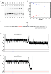
Phi29 DNAP-catalyzed replication up to or through a specific template position. (a) Time course of primer extension for a DNA hairpin substrate in bulk phase, in the presence of phi29 DNAP and 100 µM each dGTP, dCTP, dTTP and dATP. A 67 mer, 5′-6-FAM, 3′-H 14 bp hairpin (1 µM) was incubated at room temperature with 0.75 µM phi29 DNAP in buffer containing 1 mM EDTA, absent (lane 1) or present (lanes 2–7) 10 mM MgCl2 and 100 µM each of all four dNTPs (lanes 1–10) for the times indicated. The onset of primer extension requires exonucleolytic excision of the terminal ddCMP residue preceding processive dNTP additions. Reaction products were resolved on an 18% denaturing polyacrylamide gel. (b) The fluorescence intensity of bands in the gel in panel a corresponding to the intact, unextended hairpin (blue diamonds) and the extension product (red diamonds) were quantified using ImageJ software (NIH). For each lane, the fraction of the total fluorescence for these two bands was plotted as a function of reaction time. (c) DNA hairpin substrate for nanopore replication experiments. The starting abasic configuration is 5ab(18,22). In the presence of dGTP, dCTP, dTTP and ddATP, 12 nucleotides can be added up to ddATP addition in response to the first template dTMP residue (blue). This dTMP residue is positioned such that reaching this endpoint requires replication of a segment of template during which the abasic block (red Xs) is drawn into and through the nanopore lumen. After ddATP incorporation, a phi29 DNAP-DNA-dTTP ternary complex can be formed with abasic configuration 5ab(6,10). In the presence of dGTP, dCTP, dTTP and dATP, replication can proceed past the +12 position up to the abasic block. (d) phi29 DNAP-catalyzed replication on the hairpin substrate shown in panel c in the presence of 100 µM each dGTP, dCTP, dTTP and ddATP, in buffer containing 0.3 M KCl and 10 mM MgCl2. (e) phi29 DNAP-catalyzed replication after 200 µM dATP was added to the experiment shown in panel (d). Events shown in panels d and e are representative of dozens of complexes captured. Events in a control experiment in which 100 µM each dGTP, dCTP, dTTP and dATP were added absent ddATP were identical to the representative event shown in panel e. Complexes were captured within the first 10 minutes after the addition of MgCl2 to the nanopore chamber.
Figure 6
Figure 6
Phi29 DNAP-catalyzed replication by complexes held atop the nanopore at different voltages. (a) DNA hairpin substrate for nanopore replication experiments. The starting abasic configuration for this substrate is 5ab(25,29). After the exonucleolytic excision of the terminal ddCMP residue that is required for initiation of DNA synthesis, 25 nucleotides can be added before the enzyme reaches the abasic block. During DNA synthesis, the 5 abasic insert will be drawn through and past abasic configurations 5ab(18,22) to 5ab(6,10), which spans the positions mapped in Figure 3. (b) Representative current traces showing phi29 DNAP replication of the hairpin substrate shown in panel a, in buffer containing 0.3 M KCl, 10 mM MgCl2, in the presence of 100 µM each dGTP, dCTP, dTTP and dATP. Traces are shown for synthesis at (i) 220 mV, (ii) 180 mV, (iii) 140 mV, and (iv) 100 mV applied potential. Synthesis was examined within the first 10 minutes after the addition of MgCl2 to the nanopore chamber. The blue arrows below the 220 mV trace indicate the starting and end states used to quantify the synthesis rate at 220 and 100 mV.
Figure 7
Figure 7
Processive DNA replication catalyzed by phi29 DNAP on the nanopore. (a) DNA hairpin substrate for nanopore replication experiments. The starting abasic configuration for this substrate is 5ab(50,54). After the exonucleolytic excision of the terminal ddCMP residue that is required prior to DNA synthesis, 50 nucleotides can be added before the enzyme reaches the abasic block (indicated by the blue arrow above the template strand sequence). During DNA synthesis, the 5 abasic insert is drawn toward the pore lumen as the first 32 nucleotides are incorporated and the abasic configuration 5ab(18,22) is reached; subsequent nucleotide additions then draw the block up to and past configuration 5ab(6,10). Thus the abasic configurations in the amplitude map in Figure 3 are spanned. (b) Representative current trace at 180 mV applied potential showing phi29 DNAP replication of the hairpin substrate shown in panel a, in buffer containing 0.3 M KCl. (c) Representative current trace at 180 mV applied potential showing phi29 DNAP replication of the hairpin substrate shown in panel a, in buffer containing 0.6 M KCl. In panels b and c, the left and right blue arrows indicate the start and end points, respectively, used to approximate the time required to replicate ~ 50 nts along this template. Synthesis reactions were carried out in the presence of 100 µM each dGTP, dCTP, dTTP and dATP, and were examined within the first 10 minutes after the addition of MgCl2 to the nanopore chamber.

References

    1. Kapanidis AN, Strick T. Trends. Biochem. Sci. 2009;34:234–243. - PubMed
    1. Moffitt JR, Chemla YR, Smith SB, Bustamante C. Annu. Rev. Biochem. 2008;77:205–228. - PubMed
    1. Myong S, Ha T. Curr. Opin. Struct. Biol. 2010;20:121–127. - PMC - PubMed
    1. Eid J, et al. Science. 2009;323:133–138. - PubMed
    1. Harris TD, et al. Science. 2008;320:106–109. - PubMed

Publication types