. 2011 Feb;92(1):57-65.
doi: 10.1111/j.1365-2613.2010.00750.x. Epub 2010 Dec 1.

Increased connective tissue growth factor associated with cardiac fibrosis in the mdx mouse model of dystrophic cardiomyopathy

Affiliations

Increased connective tissue growth factor associated with cardiac fibrosis in the mdx mouse model of dystrophic cardiomyopathy

Carol G Au et al. Int J Exp Pathol. 2011 Feb.

Abstract

Cardiomyopathy contributes to morbidity and mortality in Duchenne muscular dystrophy (DMD), a progressive muscle-wasting disorder. A major feature of the hearts of DMD patients and the mdx mouse model of the disease is cardiac fibrosis. Connective tissue growth factor (CTGF) is involved in the fibrotic process in many organs. This study utilized the mdx mouse model to assess the role of CTGF and other extracellular matrix components during the development of fibrosis in the dystrophic heart. Left ventricular function of mdx and control mice at 6, 29 and 43 weeks was measured by echocardiography. Young (6 weeks old) mdx hearts had normal function and histology. At 29 weeks of age, mdx mice developed cardiac fibrosis and increased collagen expression. The onset of fibrosis was associated with increased CTGF transcript and protein expression. Increased intensity of CTGF immunostaining was localized to fibrotic areas in mdx hearts. The upregulation of CTGF was also concurrent with increased expression of tissue inhibitor of matrix metalloproteinases (TIMP-1). These changes persisted in 43 week old mdx hearts and were combined with impaired cardiac function and increased gene expression of transforming growth factor (TGF)-β1 and matrix metalloproteinases (MMP-2, MMP-9). In summary, an association was observed between cardiac fibrosis and increased CTGF expression in the mdx mouse heart. CTGF may be a key mediator of early and persistent fibrosis in dystrophic cardiomyopathy.


Figures

Figure 1
Figure 1
Development of cardiac fibrosis in mdx mice with age.(a–f) Masson’s trichrome stained collagen fibrils blue in myocardial sections of control C57 (a–c) and mdx (d–f) mouse hearts at ages 6 weeks (a,d), 29 weeks (b,e) and 43 weeks (c,f). All images captured at the same magnification. Scalebar represents 100 μm. (g) Area of fibrosis in histological sections was quantitated. No fibrotic areas were observed in 6 week old mice. By 29 weeks, mdx hearts displayed areas of fibrosis which persisted and were more widespread in 43 week old mdx hearts when compared to age-matched controls. (h,i) Real-time qRT-PCR analysis of mdx and control C57 mouse hearts for mRNA expression of procollagen type I (h) and procollagen type III (i). Compared to age-matched controls, expression of both procollagen type I and type III in mdx hearts was reduced at 6 weeks of age, but was increased at 29 and 43 weeks of age. The number of mice (n) analyzed for each group is listed in Table 2. Data is expressed as mean ± SEM.
Figure 2
Figure 2
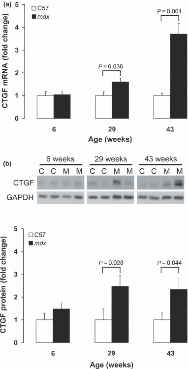
Increased connective tissue growth factor (CTGF) expression in mdx hearts. (a) CTGF mRNA levels in mdx and control C57 mice at various ages as measured by real-time qRT-PCR. Increased CTGF mRNA expression was observed in 29 week old mdx hearts, and continued to be upregulated in 43 week old mdx hearts compared to age-matched controls. (b) Representative Western blot of CTGF protein expression in control C57 (C) and mdx (M) mouse hearts (40 μg protein per lane). CTGF protein levels were quantified by densitometry scanning and adjusted for GAPDH levels. Increased CTGF protein levels were observed in mdx hearts at ages 29 and 43 weeks when compared to age-matched controls. The number of mice (n) analyzed for each group is listed in Table 2. Data is expressed as mean ± SEM.
Figure 3
Figure 3
Immunohistochemical staining for connective tissue growth factor (CTGF) in mdx hearts. Myocardial sections of control C57 (a–c) and mdx mouse (d–f) hearts at ages 6 weeks (a,d), 29 weeks (b,e) and 43 weeks (c,f) were stained with an antibody against CTGF. Positive staining shown in brown. Increased intensity of CTGF staining was noted in mdx hearts at 29 weeks and 43 weeks of age, with the strongest staining observed in fibrotic areas (arrows). All images captured at the same magnification. Scalebar represents 100 μm.
Figure 4
Figure 4
Temporal changes in transforming growth factor-β1 (TGF-β1), tissue inhibitor of matrix metalloproteinases (TIMP-1) and matrix metalloproteinases (MMP-2, MMP-9) transcript expression in mdx hearts. Real-time qRT-PCR analysis of control C57 and mdx mouse hearts at various ages for expression of (a) TGF-β1, (b) TIMP-1, (c) MMP-2 and (d) MMP-9. (a) Myocardial expression of TGF-β1 mRNA was elevated in 43 week old mdx mice compared to age-matched controls. (b) TIMP-1 expression was increased in 29 week old mdx hearts, and continued to be elevated in 43 week old mdx hearts compared to age-matched controls. (c,d) Compared to age-matched controls, myocardial expression of MMP-2 and MMP-9 was increased in 43 week old mdx mice. The number of mice (n) analyzed for each group is listed in Table 2. Data is expressed as mean ± SEM.

References

    1. Abreu JG, Ketpura NI, Reversade B, De Robertis EM. Connective-tissue growth factor (CTGF) modulates cell signalling by BMP and TGF-beta. Nat. Cell Biol. 2002;4:599–604. - PMC - PubMed
    1. Bauer R, Straub V, Blain A, Bushby K, MacGowan GA. Contrasting effects of steroids and angiotensin-converting-enzyme inhibitors in a mouse model of dystrophin-deficient cardiomyopathy. Eur. J. Heart Fail. 2009;11:463–471. - PubMed
    1. Bia BL, Cassidy PJ, Young ME, et al. Decreased myocardial nNOS, increased iNOS and abnormal ECGs in mouse models of Duchenne muscular dystrophy. J. Mol. Cell. Cardiol. 1999;31:1857–1862. - PubMed
    1. Bostick B, Yue Y, Long C, Duan D. Prevention of dystrophin-deficient cardiomyopathy in twenty-one-month-old carrier mice by mosaic dystrophin expression or complementary dystrophin/utrophin expression. Circ. Res. 2008;102:121–130. - PubMed
    1. Bushby K, Muntoni F, Bourke JP. 107th ENMC international workshop: the management of cardiac involvement in muscular dystrophy and myotonic dystrophy. 7th-9th June 2002, Naarden, the Netherlands. Neuromuscul. Disord. 2003;13:166–172. - PubMed

Publication types

Substances