. 2010 Dec 1:11:47.
doi: 10.1186/1471-2091-11-47.

Identification and characterization of a bacterial glutamic peptidase

Affiliations

Identification and characterization of a bacterial glutamic peptidase

Kenneth Jensen et al. BMC Biochem. .

Abstract

Background: Glutamic peptidases, from the MEROPS family G1, are a distinct group of peptidases characterized by a catalytic dyad consisting of a glutamate and a glutamine residue, optimal activity at acidic pH and insensitivity towards the microbial derived protease inhibitor, pepstatin. Previously, only glutamic peptidases derived from filamentous fungi have been characterized.

Results: We report the first characterization of a bacterial glutamic peptidase (pepG1), derived from the thermoacidophilic bacteria Alicyclobacillus sp. DSM 15716. The amino acid sequence identity between pepG1 and known fungal glutamic peptidases is only 24-30% but homology modeling, the presence of the glutamate/glutamine catalytic dyad and a number of highly conserved motifs strongly support the inclusion of pepG1 as a glutamic peptidase. Phylogenetic analysis places pepG1 and other putative bacterial and archaeal glutamic peptidases in a cluster separate from the fungal glutamic peptidases, indicating a divergent and independent evolution of bacterial and fungal glutamic peptidases. Purification of pepG1, heterologously expressed in Bacillus subtilis, was performed using hydrophobic interaction chromatography and ion exchange chromatography. The purified peptidase was characterized with respect to its physical properties. Temperature and pH optimums were found to be 60°C and pH 3-4, in agreement with the values observed for the fungal members of family G1. In addition, pepG1 was found to be pepstatin-insensitive, a characteristic signature of glutamic peptidases.

Conclusions: Based on the obtained results, we suggest that pepG1 can be added to the MEROPS family G1 as the first characterized bacterial member.


Figures

Figure 1
Figure 1
Phylogenetic tree of peptidases from the MEROPS peptidase family G1. The archaeal G1 peptidases are highlighted in rose. The fungal G1 peptidase cluster is highlighted in different shades of blue, and the major fungal clusters are labeled A, B and C. All annotated and putative family G1 peptidases (except for non-peptidase homologs) at the MEROPS peptidase database (version 9.1) were aligned using ClustalX version 2.0.11. The bootstrapped maximum likelihood tree was built using PhyML 3.0 aLRT [31] and visualized in TreeView [32]. The tree was bootstrapped with 100 iterations and bootstrap values are indicated on the figure. All GenBank accession numbers and detailed information on the members of each cluster can be found in Additional file 1 Table S1. Asa: [GenBank: YP_003816089] from Acidilobus saccharovorans; Ame: [GenBank: YP_003762485] from Amycolatopsis mediterranei; Bin: [GenBank: ACB95479] from Beijerinckia indica; Bvi: [GenBank: ABO59772] from Burkholderia vietnamiensis; Cat: [GenBank: YP_003114490] from Catenulispora acidiphila; Ckl: [GenBank: BAH07727] from Clostridium kluyveri; Cma: [GenBank: ABW02092] from Caldivirga maquilingensis; Geo: [GenBank: YP_003244752] from Geobacillus sp. Y412MC10; PepG1: [GenBank: HM011103] from Alicyclobacillus sp. DSM 15716; Rsa_1: [GenBank: ABY24309] from Renibacterium salmoninarum; Rsa_2: [GenBank: ABY21885] from Renibacterium salmoninarum.
Figure 2
Figure 2
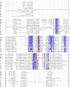
Comparison of pepG1 with well-known family G1 peptidase and putative bacterial and archaeal members. Full-length sequences including signal peptides were aligned using ClustalX version 2.0.11. The residues numbering for each peptidase is indicated. The seven highly conserved segments in all G1 peptidases are colored according to the percentage of the residues in each column that agrees with the consensus sequence. Only the residues that agree with the consensus residue for each column are colored. Dark blue means > 80%, blue > 60%, light blue > 40% and white < 40%. The catalytic dyad is colored red and the residues involved in a highly conserved disulfide bridge are shown in yellow. The fungal peptidases used for the alignment were aspergilloglutamic peptidase (AGP, [GenBank: P24665]) from Aspergillus niger, scytalidoglutamic peptidase (SGP, [GenBank: P15369]) from Scytalidium lignicolum, acid peptidases B and C (EapB [GenBank: Q00550] and EapC [GenBank: Q00551]) from Cryphonectria parisitica, Penicillium marneffei acid proteinase (PMAP-1, [GenBank: EEA28697]), BcACP1 ([GenBank: AAZ77775) from Botryotinia fuckeliana and Talaromyces emersonii glutamic peptidase 1 (TGP1, [GenBank: Q8X1C6]). The putative bacterial peptidases were [GenBank: YP_003762485] from Amycolatopsis mediterranei (Ame), [GenBank: ACB95479] from Beijerinckia indica (Bin), [GenBank: ABO59772] from Burkholderia vietnamiensis (Bvi), [GenBank: YP_003114490] from Catenulispora acidiphila (Cat), [GenBank: BAH07727] from Clostridium kluyveri (Ckl) and [GenBank: ABY24309], [GenBank: YP_003244752] from Geobacillus sp. (Geo), [GenBank: HM011103] from Alicyclobacillus sp. DSM 15716 (pepG1) and [GenBank: ABY21885] from Renibacterium salmoninarum (Rsa_1 and Rsa_2). The two archaeal peptidases were [GenBank: YP_003816089] from Acidilobus saccharovorans (Asa) and [GenBank: ABW02092] from Caldivirga maquilingensis (Cma).
Figure 3
Figure 3
Homology model of pepG1. The model was generated using SWISS-MODEL [18] and visualized using PYMOL. The active site residues, Q117 and E199, are shown in yellow. The upper antiparallel β-sheet is light blue, and the lower β-sheet is red.
Figure 4
Figure 4
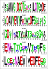
WebLogo of the protein signature PR00977. The sequence logo was constructed from the alignment of the four G1 peptidases AGP, SGP, EapB and EapC [22]. The letter size is proportional to the degree of amino acid conservation. The WebLogo was generated using WebLogo version 2.8.2 [34].
Figure 5
Figure 5
Purification of pepG1. Fractions 49-53 were subjected to SDS-page (insert). The size of the molecular marker is indicated on the left side of the SDS-page gel. The SDS-page gel was stained with Coomasie Brilliant Blue. The bars under the A280 trace indicate the activity of the individual fractions towards Protazyme OL (crosslinked and dyed collagen) at pH 4.0, 37°C.
Figure 6
Figure 6
Characterization of pepG1. A. Effect of pH on pepG1 activity. The maximum activity at 37°C towards AZCL-collagen was obtained at a broad plateau around pH 3.0 and set at 100%. B. Determination of temperature optimum for pepG1. The maximum activity towards AZCL-collagen was observed at 60°C, pH 4.0 and set at 100%. C. pH stability of pepG1. pepG1 was diluted and incubated in assay buffer pH 2-12 for two hours at 37°C. pH was then adjusted to pH 4.0 and activity was measured at 37°C. D. Temperature stability of pepG1. pepG1 was incubated at 50°C (black), 60°C (grey) and 70°C (light grey) for up to one hour, cooled to 4°C on ice and assayed at 37°C, pH 4.0. Stability is measured relative to samples incubated on ice.

References

    1. Becker F, Schnorr K, Wilting R, Tolstrup N, Bendtsen JD, Olsen PB. Development of in vitro transposon assisted signal sequence trapping and its use in screening Bacillus halodurans C125 and Sulfolobus solfataricus P2 gene libraries. J Microbiol Methods. 2004;57(1):123–133. doi: 10.1016/j.mimet.2003.12.002. - DOI - PubMed
    1. Rawlings ND, Morton FR, Kok CY, Kong J, Barrett AJ. MEROPS: the peptidase database. Nucleic Acids Res. 2008. pp. D320–325. - PMC - PubMed
    1. Sims AH, Dunn-Coleman NS, Robson GD, Oliver SG. Glutamic protease distribution is limited to filamentous fungi. FEMS Microbiol Lett. 2004;239(1):95–101. doi: 10.1016/j.femsle.2004.08.023. - DOI - PubMed
    1. Inoue H, Kimura T, Makabe O, Takahashi K. The gene and deduced protein sequences of the zymogen of Aspergillus niger acid proteinase A. J Biol Chem. 1991;266(29):19484–19489. - PubMed
    1. Oda N, Gotoh Y, Oyama H, Murao S, Oda K, Tsuru D. Nucleotide sequence of the gene encoding the precursor protein of pepstatin insensitive acid protease B, scytalidopepsin B, from Scytalidium lignicolum. Biosci Biotechnol Biochem. 1998;62(8):1637–1639. doi: 10.1271/bbb.62.1637. - DOI - PubMed

Publication types

MeSH terms

LinkOut - more resources