

Quantitative shear wave ultrasound elastography: initial experience in solid breast masses
- PMID: 21122101
- PMCID: PMC3046449
- DOI: 10.1186/bcr2787
Quantitative shear wave ultrasound elastography: initial experience in solid breast masses
Abstract
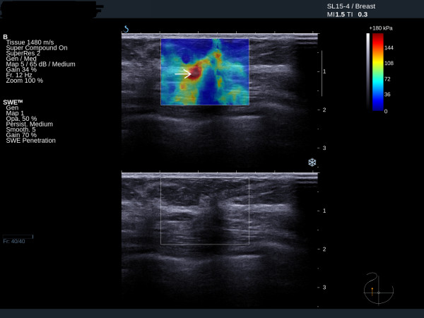
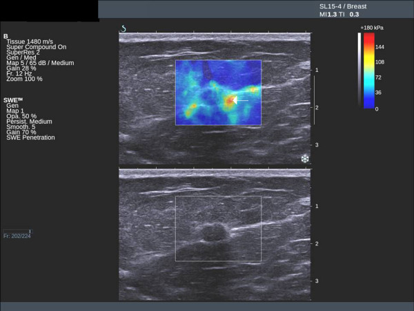
Introduction: Shear wave elastography is a new method of obtaining quantitative tissue elasticity data during breast ultrasound examinations. The aims of this study were (1) to determine the reproducibility of shear wave elastography (2) to correlate the elasticity values of a series of solid breast masses with histological findings and (3) to compare shear wave elastography with greyscale ultrasound for benign/malignant classification.
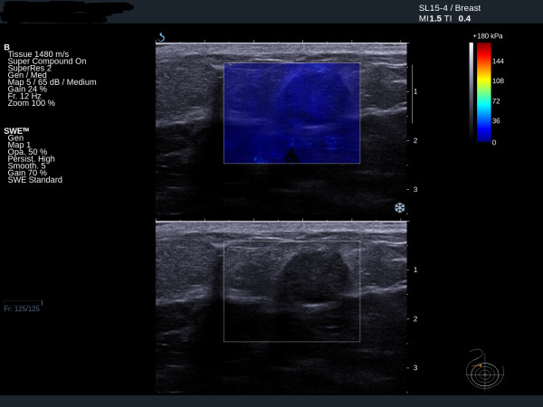
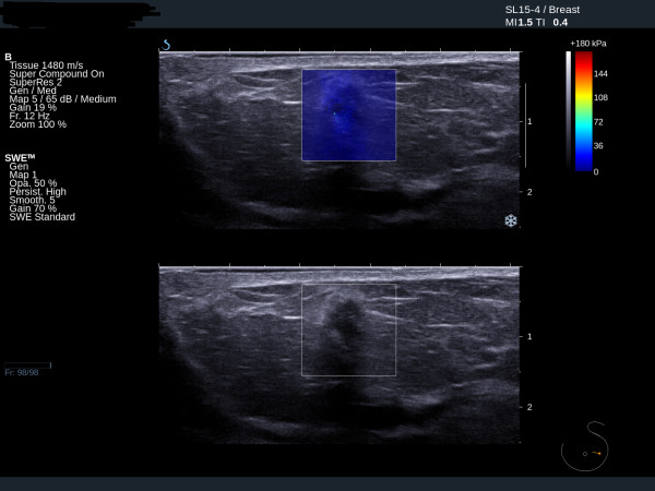
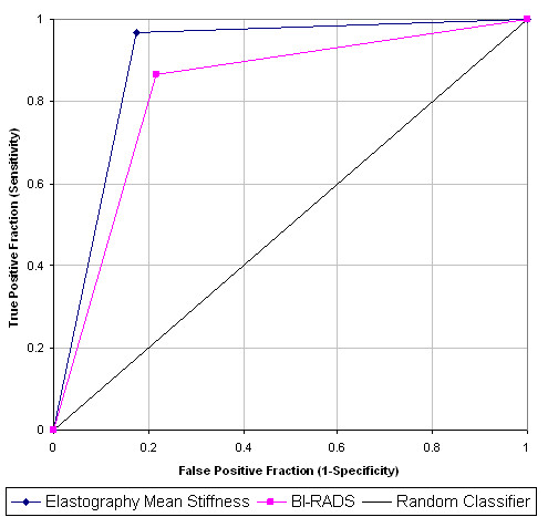
Methods: Using the Aixplorer® ultrasound system (SuperSonic Imagine, Aix en Provence, France), 53 solid breast lesions were identified in 52 consecutive patients. Two orthogonal elastography images were obtained of each lesion. Observers noted the mean elasticity values in regions of interest (ROI) placed over the stiffest areas on the two elastography images and a mean value was calculated for each lesion. A sub-set of 15 patients had two elastography images obtained by an additional operator. Reproducibility of observations was assessed between (1) two observers analysing the same pair of images and (2) findings from two pairs of images of the same lesion taken by two different operators. All lesions were subjected to percutaneous biopsy. Elastography measurements were correlated with histology results. After preliminary experience with 10 patients a mean elasticity cut off value of 50 kilopascals (kPa) was selected for benign/malignant differentiation. Greyscale images were classified according to the American College of Radiology (ACR) Breast Imaging Reporting and Data System (BI-RADS). BI-RADS categories 1-3 were taken as benign while BI-RADS categories 4 and 5 were classified as malignant.
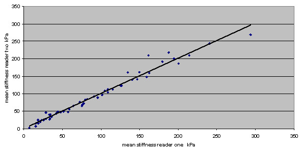
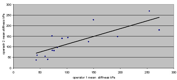
Results: Twenty-three benign lesions and 30 cancers were diagnosed on histology. Measurement of mean elasticity yielded an intraclass correlation coefficient of 0.99 for two observers assessing the same pairs of elastography images. Analysis of images taken by two independent operators gave an intraclass correlation coefficient of 0.80. Shear wave elastography versus greyscale BI-RADS performance figures were sensitivity: 97% vs 87%, specificity: 83% vs 78%, positive predictive value (PPV): 88% vs 84%, negative predictive value (NPV): 95% vs 82% and accuracy: 91% vs 83% respectively. These differences were not statistically significant.
Conclusions: Shear wave elastography gives quantitative and reproducible information on solid breast lesions with diagnostic accuracy at least as good as greyscale ultrasound with BI-RADS classification.
Figures
References
-
- Zonderland HM, Coerkamp EG, Hermans J, Van De Vijver MJ, Van Voorthuisen AE. Diagnosis of breast cancer: contribution of US as an adjunct to mammography. Radiology. 1999;213:413–422. - PubMed
-
- Stavros AT, Thickman D, Rapp CL, Dennis MA, Parker SH, Sisney GA. Solid breast nodules: use of sonography to distinguish between benign and malignant lesions. Radiology. 1995;196:123–134. - PubMed
-
- Fasching PA, Heusinger K, Loehberg CR, Wenkel E, Lux MP, Schrauder M, Koscheck T, Bautz W, Schulz-Wendtland R, Beckmann MW, Bani MR. Influence of mammographic density on the diagnostic accuracy of tumor size assessment and association with breast cancer tumor characteristics. Eur J Radiol. 2006;60:398–404. doi: 10.1016/j.ejrad.2006.08.002. - DOI - PubMed
-
- Parker SH, Jobe WE, Dennis MA, Stavros AT, Johnson KK, Yakes WF, Truell JE, Price JG, Kortz AB, Clark DG. US-guided automated large-core breast biopsy. Radiology. 1993;187:507–511. - PubMed
MeSH terms
LinkOut - more resources
Full Text Sources
Other Literature Sources
Medical
Research Materials
