. 2010 Dec 1:10:308.
doi: 10.1186/1471-2180-10-308.

Bacteriocyte dynamics during development of a holometabolous insect, the carpenter ant Camponotus floridanus

Affiliations

Bacteriocyte dynamics during development of a holometabolous insect, the carpenter ant Camponotus floridanus

Sascha Stoll et al. BMC Microbiol. .

Abstract

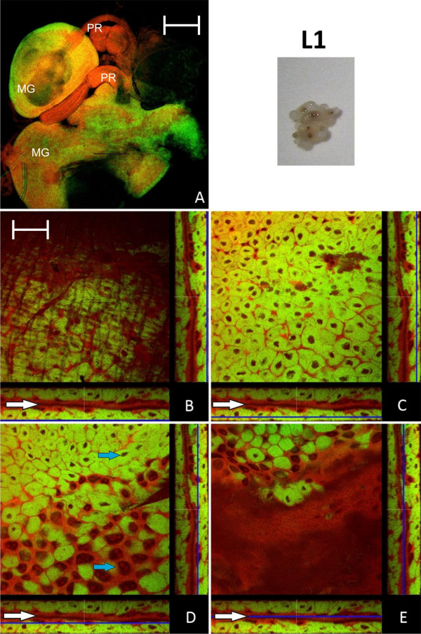
Background: The carpenter ant Camponotus floridanus harbors obligate intracellular mutualistic bacteria (Blochmannia floridanus) in specialized cells, the bacteriocytes, intercalated in their midgut tissue. The diffuse distribution of bacteriocytes over the midgut tissue is in contrast to many other insects carrying endosymbionts in specialized tissues which are often connected to the midgut but form a distinct organ, the bacteriome. C. floridanus is a holometabolous insect which undergoes a complete metamorphosis. During pupal stages a complete restructuring of the inner organs including the digestive tract takes place. So far, nothing was known about maintenance of endosymbionts during this life stage of a holometabolous insect. It was shown previously that the number of Blochmannia increases strongly during metamorphosis. This implicates an important function of Blochmannia in this developmental phase during which the animals are metabolically very active but do not have access to external food resources. Previous experiments have shown a nutritional contribution of the bacteria to host metabolism by production of essential amino acids and urease-mediated nitrogen recycling. In adult hosts the symbiosis appears to degenerate with increasing age of the animals.

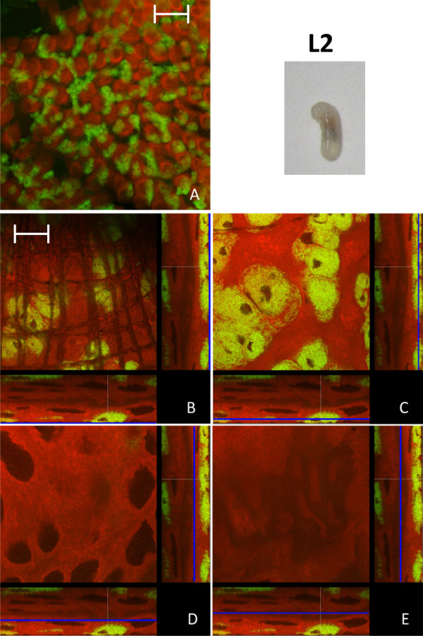
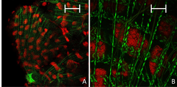
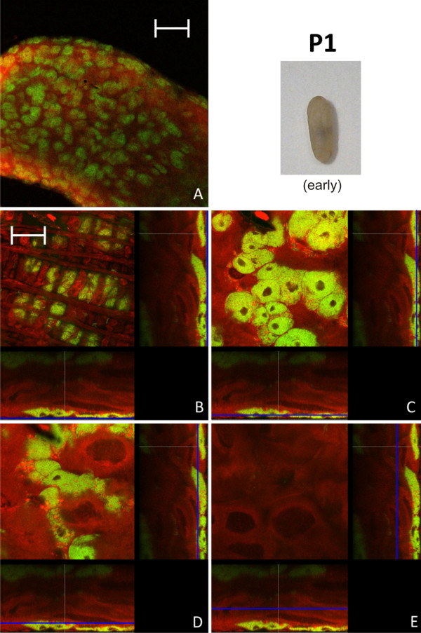
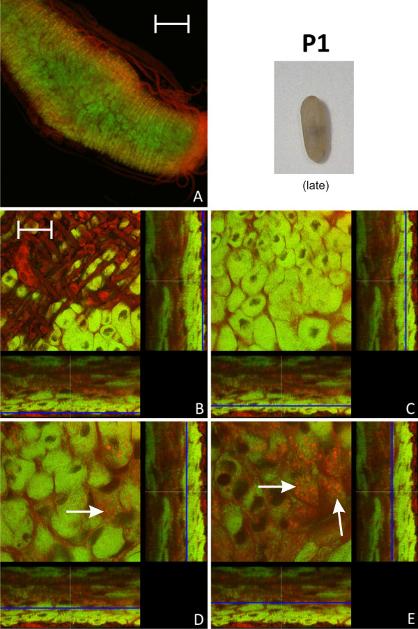
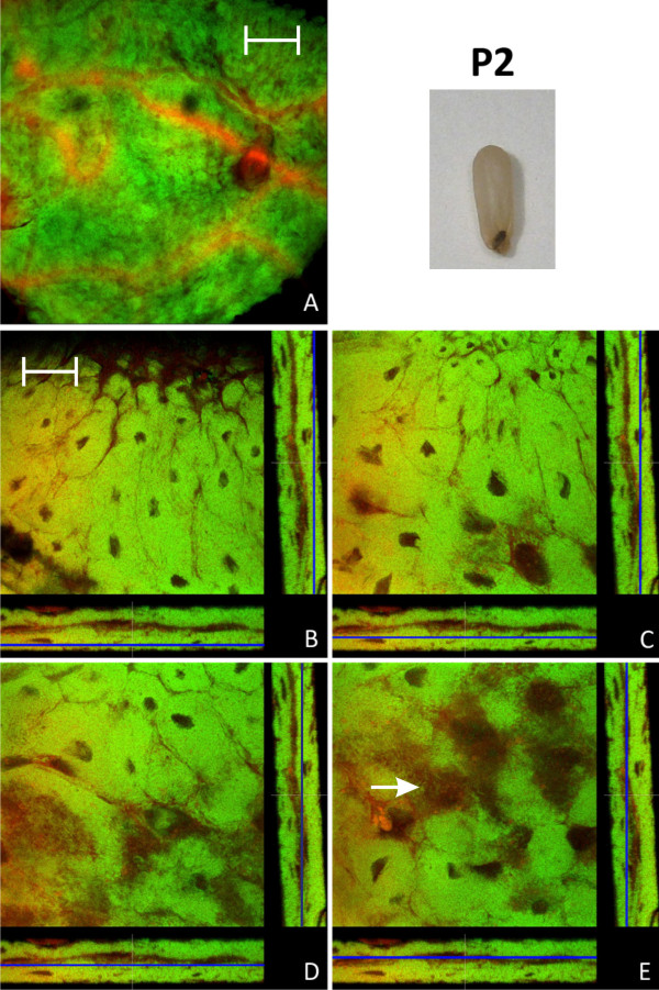
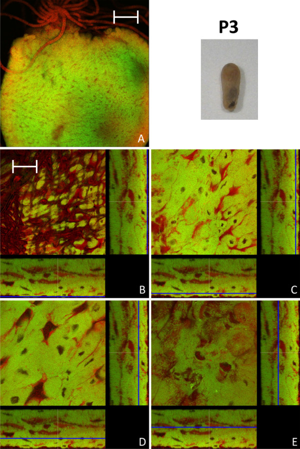
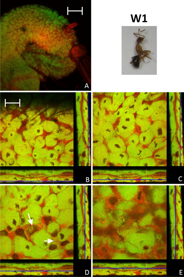
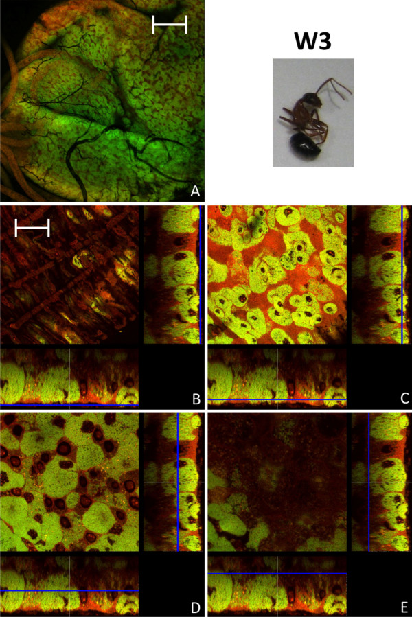
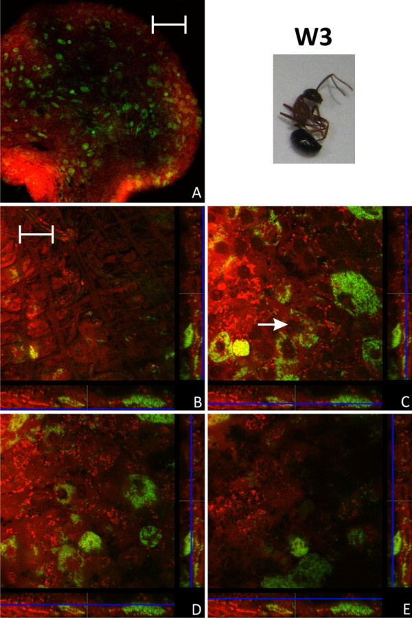
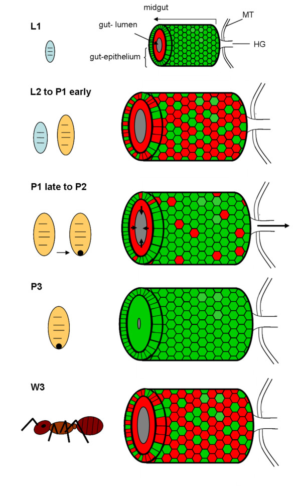
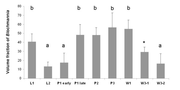
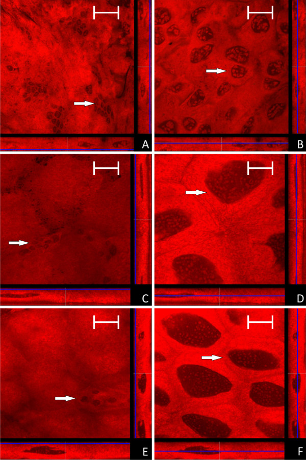
Results: We investigated the distribution and dynamics of endosymbiotic bacteria and bacteriocytes at different stages during development of the animals from larva to imago by confocal laser scanning microscopy. The number of bacteriocytes in relation to symbiont-free midgut cells varied strongly over different developmental stages. Especially during metamorphosis the relative number of bacteria-filled bacteriocytes increased strongly when the larval midgut epithelium is shed. During this developmental stage the midgut itself became a huge symbiotic organ consisting almost exclusively of cells harboring bacteria. In fact, during this phase some bacteria were also found in midgut cells other than bacteriocytes indicating a cell-invasive capacity of Blochmannia. In adult animals the number of bacteriocytes generally decreased.

Conclusions: During the life cycle of the animals the distribution of bacteriocytes and of Blochmannia endosymbionts is remarkably dynamic. Our data show how the endosymbiont is retained within the midgut tissue during metamorphosis thereby ensuring the maintenance of the intracellular endosymbiosis despite a massive reorganization of the midgut tissue. The transformation of the entire midgut into a symbiotic organ during pupal stages underscores the important role of Blochmannia for its host in particular during metamorphosis.


Figures

Figure 1
Figure 1
Larva of stage L1. A: Overview showing two midguts (MG) and their proventriculi (PR) by confocal laser scanning microscopy. B - E: Four orthogonal views of confocal image stacks of C floridanus L1 larva midgut sections. The blue lines in the XZ and YZ stack representations (below and on the right side of each quadratic micrograph) illustrate the position of the image plane (XY). The bacteria-free midgut cells typically have large nuclei and several nucleoli while the bacteriocytes are characterized by small nuclei (blue arrows in D). The bacteriocytes form a nearly contiguous layer surrounding the midgut (B, C) directly underneath of the muscle network (A and Fig. 3). There are no bacteriocytes present in the cell layer lining the midgut lumen (D, E). The midgut lumen is indicated by white arrows. Green label: The Blochmannia specific probe Bfl172-FITC; red label: SYTO Orange 83. The scale bars correspond to 220 μM (A) and 35 μM (B - E), respectively.
Figure 2
Figure 2
Larva of stage L2. Overview (A) and detailed images of different optical sections (B - E) of the midgut of a C. floridanus larva (L2) by confocal laser scanning microscopy (for further information regarding the composition of the figure see legend of Fig. 1). The bacteriocytes are located in cell clusters of different size on the outer surface of the midgut (B, C) and the cells lining the midgut lumen are free of bacteria (D, E). Green label: The Blochmannia specific probe Bfl172-FITC; red label: SYTO Orange 83. The scale bars correspond to 220 μM (A) and 35 μM (B - E), respectively.
Figure 3
Figure 3
Overview (A) and detailed image (B) of the actin-stained muscle network surrounding the midgut of a B. floridanus larva (L2) by confocal laser scanning microscopy. Green label: FITC-Phalloidin; red label: The Blochmannia specific probe Bfl172-Cy3. The scale bars correspond to 220 μM (A) and 35 μM (B), respectively.
Figure 4
Figure 4
Early stage P1 pupa. Overview (A) and detailed images of different optical sections (B - E) of the midgut of a C. floridanus pupa (P1) prior to excretion of the meconium by confocal laser scanning microscopy (for further information regarding the composition of the figure see legend of Fig. 1). The distribution of bacteriocytes resembles that of L2 larvae shown in Fig. 2. Green label: The Blochmannia specific probe Bfl172-FITC; red label: SYTO Orange 83. The scale bars correspond to 220 μM (A) and 35 μM (B - E), respectively.
Figure 5
Figure 5
Late stage P1 pupa. Overview (A) and detailed images of different optical sections (B - E) of the midgut of a C. floridanus pupa (P1) at a later stage than the pupa shown in Fig. 4 by confocal laser scanning microscopy (for further information regarding the composition of the figure see legend of Fig. 1). The bacteriocyte layer encloses the entire midgut (C) and the infection of midgut cells other than bacteriocytes (i.e. cells with large and nucleoli-rich nuclei) is increasingly observed (white arrows in D, E). Bacteria-harboring cells are now found in the epithelial layer bordering the gut lumen (E). Green label: The Blochmannia specific probe Bfl172-FITC; red label: SYTO Orange 83. The scale bars correspond to 220 μM (A) and 35 μM (B - E), respectively.
Figure 6
Figure 6
Pupa of stage P2. Overview (A) and detailed images of different optical sections (B - E) of the midgut of a pupa after excretion of the meconium (P2) by confocal laser scanning microscopy (for further information regarding the composition of the figure see legend of Fig. 1). Virtually all cells of the midgut harbor Blochmannia and the bacteria once more are present in cells with large and nucleoli-rich nuclei (e.g. white arrow in figure part E). Green label: The Blochmannia specific probe Bfl172-FITC; red label: SYTO Orange 83. The scale bars correspond to 220 μM (A) and 35 μM (B - E), respectively.
Figure 7
Figure 7
Pupa of stage P3. Overview (A, red stained Malpighian tubules are visible on the top of the midgut) and detailed images of different optical sections (B - E) of the midgut of a pupa immediately before eclosion (P3) by confocal laser scanning microscopy (for further information regarding the composition of the figure see legend of Fig. 1). In comparison to P2 (see Fig. 6), a slight increase in bacteria-free midgut cells with large nuclei can be observed. Green label: The Blochmannia specific probe Bfl172-FITC; red label: SYTO Orange 83. The scale bars correspond to 220 μM (A) and 35 μM (B - E), respectively.
Figure 8
Figure 8
Imago of stage W1. Overview (A) and detailed images of different optical sections (B - E) of the midgut of a young worker shortly after eclosion (W1) by confocal laser scanning microscopy (for further information regarding the composition of the figure see legend of Fig. 1). In the overview (A) the proventriculus can be seen on the right side of the midgut. The number of not-infected cells with larger nuclei is increased in comparison to the late pupae stages (Fig. 7). Still there are bacteria in cells which do not resemble typical bacteriocytes (e.g. white arrows in figure part D). Green label: The Blochmannia specific probe Bfl172-FITC; red label: SYTO Orange 83. The scale bars correspond to 220 μM (A) and 35 μM (B - E), respectively.
Figure 9
Figure 9
Imago of stage W3. Overview (A) and detailed images of different optical sections (B - E) of the midgut of a worker several months of age (W3) by confocal laser scanning microscopy (for further information regarding the composition of the figure see legend of Fig. 1). The proportion of bacteria-free cells is strongly increased, but still there are many bacteriocytes present. Green label: The Blochmannia specific probe Bfl172-FITC; red label: SYTO Orange 83. The scale bars correspond to 220 μM (A) and 35 μM (B - E), respectively.
Figure 10
Figure 10
Imago of stage W3. Overview (A) and detailed images of different optical sections (B - E) of the midgut of another worker several months of age (W3) by confocal laser scanning microscopy (for further information regarding the composition of the figure see legend of Fig. 1). The number of bacteriocytes is strongly reduced as compared to the worker (W3) shown in Fig. 9. Bacteria present in other cell types than bacteriocytes can be observed (e.g. white arrow in figure part C). Green label: The Blochmannia specific probe Bfl172-FITC; red label: SYTO Orange 83. The scale bars correspond to 220 μM (A) and 35 μM (B - E), respectively.
Figure 11
Figure 11
Schematic overview of distribution of Blochmannia in the migut epithelium during host ontogeny, summarizing results of Fig. 1 to Fig. 10. Red coloured cells are free of Blochmannia and green coloured cells are filled with endosymbionts. In small larvae (L1) all cells of the outer layer of the midgut tissue are filled with bacteria, whereas inner layers are devoid of Blochmannia. In larger larvae (L2) and pupae directly after pupation (P1 early) the midgut-epithelium strongly expands paralleling the growth of the individual. A large number of cells in the outer cell layer do not contain Blochmannia at this stage. During metamorphosis the larval gut epithelium is shed (P1 late to P2) and excreted, forming the meconium (dark spot) in the distal end of the pupal case. During this stage an increased number of cells in the outer layer of the midgut-epithelium harbour Blochmannia. In pupae directly before eclosion (P3) the circumference of the gut lumen is very tiny as it is empty. At this stage the whole midgut can be viewed as a bacteriome, since almost all cells forming the midgut-epithelium harbour Blochmannia. After eclosion of workers the symbiosis degrades. In old workers (W3) the majority of cells in the outer layer of the epithelium do not contain Blochmannia any longer and the inner layer even less so. The circumference of the gut lumen is larger again. MT: Malphigian tubules, HG: hingut.
Figure 12
Figure 12
The figure shows volume fractions of Blochmannia symbionts in the midgut tissue of the various developmental stages shown in Fig. 1 to Fig. 10 calculated from the confocal image stacks as described in the Methods section in arbitrary units. The results show the strong relative decrease of Blochmannia-bearing midgut cells between L1 and L2, the strong increase in bacteria-infected cells during the P1 stage and the decrease of bacteria-infected cells in adult animals. Standard deviations are shown as vertical bars on top of the columns. Groups differing significantly at the p < 0.05 level in a Tukey HSD post hoc test are marked with different letters above bars. * W3-1 was not included in the statistical analysis due to small sample size.
Figure 13
Figure 13
Confocal micrographs of the midgut of larvae and pupae derived from antibiotics treated queens (for further information regarding the composition of the figure see legend of Fig. 1). No Blochmannia specific signal is detectable in any of the preparations. Cells resembling empty bacteriocytes are located as small cell clusters on the outer face of the midgut (marked with a white arrow in figure's parts A, C, E), while typical epithelial cells show the characteristic large nuclei (marked with a white arrow in figure's parts B, D, F). A and B: young larvae (L1); C and D: older larvae (L2); E and F: young pupae (P1). Green label: The Blochmannia specific probe Bfl172-FITC; red label: SYTO Orange 83. The scale bar corresponds to 35 μM.

References

    1. Buchner P. Endosymbiosis of animals with plant microorganisms. Intersciences Publishers Inc. New York, N.Y; 1965.
    1. Baumann P. Biology of bacteriocyte-associated endosymbionts of plant sap-sucking insects. Annu Rev Microbiol. 2005;59:155–189. doi: 10.1146/annurev.micro.59.030804.121041. - DOI - PubMed
    1. Wernegreen JJ. Genome evolution in bacterial endosymbionts of insects. Nat Rev Genet. 2002;3:850–861. doi: 10.1038/nrg931. - DOI - PubMed
    1. Sauer C, Dudaczek D, Hölldobler B, Gross R. Tissue localization of the endosymbiotic bacterium "Candidatus Blochmannia floridanus" in adults and larvae of the carpenter ant Camponotus floridanus. Appl Environ Microbiol. 2002;68:4187–4193. doi: 10.1128/AEM.68.9.4187-4193.2002. - DOI - PMC - PubMed
    1. Schröder D, Deppisch H, Obermayer M, Krohne G, Stackebrandt E, Hölldobler B, Goebel W, Gross R. Intracellular endosymbiotic bacteria of Camponotus species (carpenter ants): systematics, evolution and ultrastructural characterization. Mol Microbiol. 1996;21:479–489. doi: 10.1111/j.1365-2958.1996.tb02557.x. - DOI - PubMed

Publication types

LinkOut - more resources