. 2010 Dec 1:10:374.
doi: 10.1186/1471-2148-10-374.

Conservation, loss, and redeployment of Wnt ligands in protostomes: implications for understanding the evolution of segment formation

Affiliations

Conservation, loss, and redeployment of Wnt ligands in protostomes: implications for understanding the evolution of segment formation

Ralf Janssen et al. BMC Evol Biol. .

Abstract

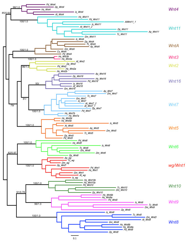
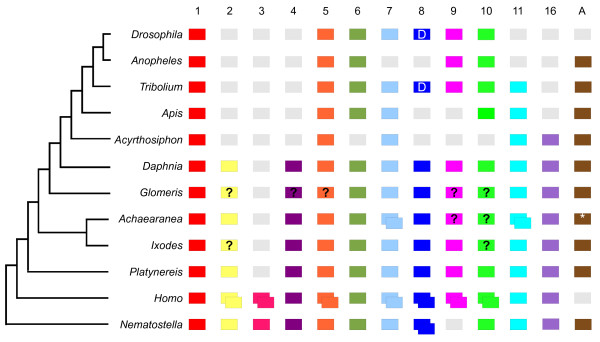
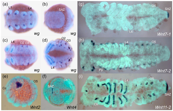
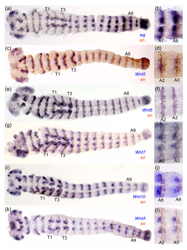
Background: The Wnt genes encode secreted glycoprotein ligands that regulate a wide range of developmental processes, including axis elongation and segmentation. There are thirteen subfamilies of Wnt genes in metazoans and this gene diversity appeared early in animal evolution. The loss of Wnt subfamilies appears to be common in insects, but little is known about the Wnt repertoire in other arthropods, and moreover the expression and function of these genes have only been investigated in a few protostomes outside the relatively Wnt-poor model species Drosophila melanogaster and Caenorhabditis elegans. To investigate the evolution of this important gene family more broadly in protostomes, we surveyed the Wnt gene diversity in the crustacean Daphnia pulex, the chelicerates Ixodes scapularis and Achaearanea tepidariorum, the myriapod Glomeris marginata and the annelid Platynereis dumerilii. We also characterised Wnt gene expression in the latter three species, and further investigated expression of these genes in the beetle Tribolium castaneum.

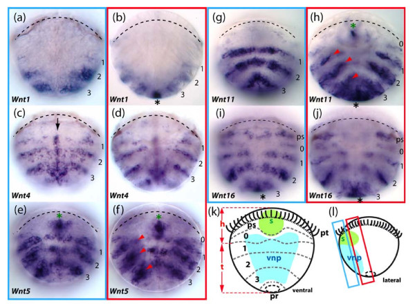
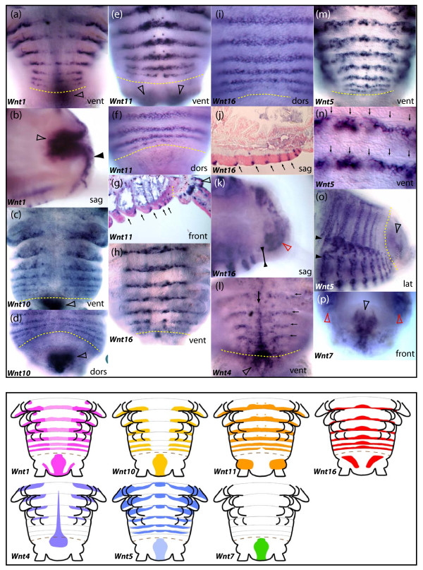
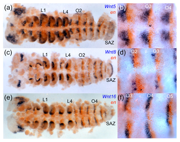
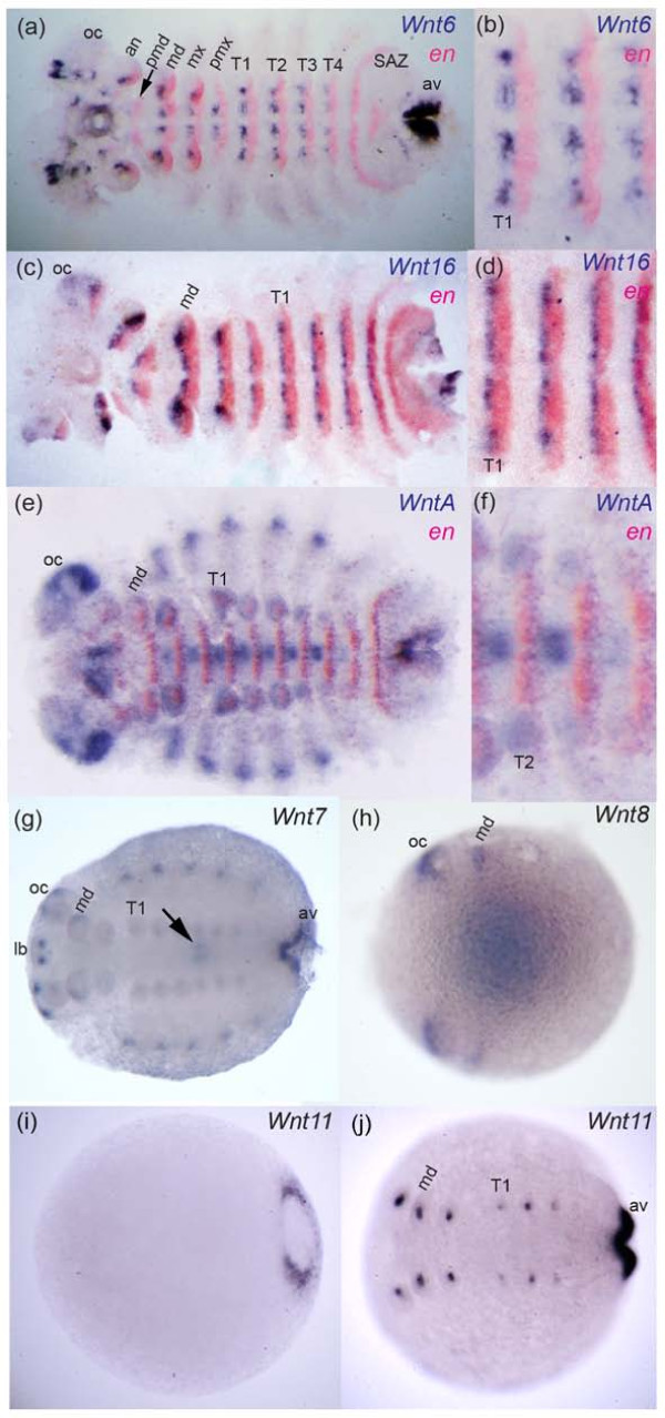
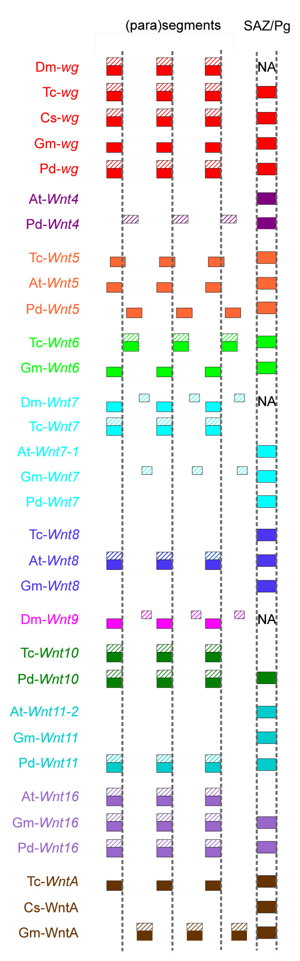
Results: We found that Daphnia and Platynereis both contain twelve Wnt subfamilies demonstrating that the common ancestors of arthropods, ecdysozoans and protostomes possessed all members of all Wnt subfamilies except Wnt3. Furthermore, although there is striking loss of Wnt genes in insects, other arthropods have maintained greater Wnt gene diversity. The expression of many Wnt genes overlap in segmentally reiterated patterns and in the segment addition zone, and while these patterns can be relatively conserved among arthropods and the annelid, there have also been changes in the expression of some Wnt genes in the course of protostome evolution. Nevertheless, our results strongly support the parasegment as the primary segmental unit in arthropods, and suggest further similarities between segmental and parasegmental regulation by Wnt genes in annelids and arthropods respectively.

Conclusions: Despite frequent losses of Wnt gene subfamilies in lineages such as insects, nematodes and leeches, most protostomes have probably maintained much of their ancestral repertoire of twelve Wnt genes. The maintenance of a large set of these ligands could be in part due to their combinatorial activity in various tissues rather than functional redundancy. The activity of such Wnt 'landscapes' as opposed to the function of individual ligands could explain the patterns of conservation and redeployment of these genes in important developmental processes across metazoans. This requires further analysis of the expression and function of these genes in a wider range of taxa.


Figures

Figure 1
Figure 1
Maximum likelihood tree of Wnt amino acid sequences in selected metazoans. Bootstrap values/poster probabilities from Maximum likelihood and Bayesian analyses respectively are given on branches. Note that support for the position of Is_Wnt16 was only found using Maximum likelihood (see also Additional file 5). Wnt amino acid sequences were used from the following species: Achaearanea tepidariorum (At), Acyrthosiphon pisum (Ap), Cupiennius salei (Cs), Daphnia pulex (Dp), Drosophila melanogaster (Dm), Glomeris marginata (Gm), Homo sapiens (Hs), Ixodes scapularis (Is), Platynereis dumerilii (Pd) and Tribolium castaneum (Tc). Bootstrap values and posterior probabilities of all branches are given in Additional files 6 and 7 respectively.
Figure 2
Figure 2
Metazoan Wnt genes. The Wnt subfamilies (1 to 11, 16 and A) found in the various metazoans are represented by coloured boxes. Grey boxes indicate the loss of particular Wnt subfamilies and boxes with question marks indicate Wnts not found in some animals, but which cannot be definitively described as 'lost' because the relevant genomes have not been sequenced or require more comprehensive annotation. Duplicated Wnts are represented by two overlapping boxes. Note that Wnt8 is also called WntD in Drosophila and Tribolium. The phylogenetic relationships of the various animals is indicated by the tree on the left [14-16,21]. The asterisk indicates that for WntA an orthologue was isolated from another spider, Cupiennius. Note that the complete Achaearanea Wnt6 sequence was only identified subsequent to the phylogenetic analysis.
Figure 3
Figure 3
wg, Wnt2, Wnt4, Wnt7-1, Wnt7-2 and Wnt11-2 expression in Achaearanea. At-wg is expressed in the anterior ventral portion of the limb buds (a), but is not observed in ventral or dorsal regions of the prosomal segments or in the SAZ (b). Expression of At-wg extends along the axis of the growing legs and is observed in dorsal stripe in O2 and O3 (c) and later in O1 to O5 in groups of cells in the opisthosomal limb buds (d). At-Wnt2 expression is observed from stage 9 onwards in a central and lateral triangular shaped domain in the developing head lobes (e). At-Wnt4 is only expressed in a few cells at the very posterior of the SAZ during late embryonic development (f). Whereas At-Wnt7-1 is only expressed in the SAZ (g), At-Wnt7-2 is expressed at the base of the appendages and in a lateral anterior and posterior domain in the head lobes (h). At-Wnt11-2 expression appears at stage 6 in the posterior end of the embryo and persists in the SAZ throughout embryonic development (i). At-Wnt11-2 is also expressed in an anterior domain along the proximo-distal leg axis, the buds of the opisthosomal appendages and in the stomodeal region (i). Ch, cheliceres; Pp, pedipalps; L1 and L4, leg bearing segments; O1 to O5, opisthosomal segments; SAZ, segment addition zone. (a) to (d), ventral views of whole mounted embryos. (e), lateral view. (f), anterior view with posterior curving to the right. (g) to (i), flat mounted embryos with anterior to the left.
Figure 4
Figure 4
Expression of wg, Wnt5, Wnt6, Wnt7 Wnt10 and WntA with respect to en in Tribolium. Germ band extended Tribolium embryos double stained for transcripts of en and wg (a), (b); Wnt5 (c), (d); Wnt6 (e), (f); Wnt7 (g), (h); Wnt10 (i), (j) and WntA (k), (l). High magnification images of segments are shown in (b), (d), (f), (h), (j) and (l). All embryos are shown with anterior to the left. thoracic (T1, T2) and abdominal (A2, A3, A8, A9) segments are indicated.
Figure 5
Figure 5
Expression patterns of five Wnt genes in 48 hours post-fertilization trochophore larvae of the annelid Platynereis. The expression of these Wnt genes is observed either in reiterated ectodermal segmental stripes and/or in the pygidial/proctodeal presumptive territory. A schematic description of the trochophore larva is given in (k). pt: the prototroch, a ciliated belt used for swimming, also highlighted by a black line on larvae photographs, divides the larva into an apical episphere and a vegetal hyposphere; pr: proctodeum; s: stomodeum; ps: peristomium, a band of embryonic tissues around the forming mouth; 0: anterior-most segmental unit; 1-3: presumptive areas of the larval appendage-bearing segments. h: the future head of the worm formed by the episphere plus peristomium plus segment 0; t: the future trunk of the worm formed by larval segments 1-3 plus the pygidium. (l) shows the two approximate focal planes that are used for larvae photographs. The first and third panel columns (a), (c), (e), (g), (i) show ventral views of trochophore larvae, focusing mainly on tissues of the ventral neuroectoderm that will form the ventral nerve cord. The second and fourth panel columns (b), (d), (f), (h), (j) are frontal optical section focusing on the lateral parapodia-forming fields. The Wnt stripes corresponding to each presumptive larval segment are numbered 1, 2, 3. In addition, a more anterior metameric unit located just below the prototroch is numbered 0. This unit does not produce a larval segment but fuses with the head early in development. Black asterisks show expressions in the pygidial/proctodeal area. Green asterisks show expressions in the stomodeal bulb that will give rise to the mouth. The midline expression of Pd-Wnt4 is indicated by a black arrow. Internal Pd-Wnt5 and Pd-Wnt11 expression potentially located either in the segmental mesoderm or in ectodermal cells of the chaetal sacs are shown by red arrowheads. Additional expression of Wnt genes in the nascent brain are described elsewhere [84].
Figure 6
Figure 6
Expression patterns of seven Wnt genes during posterior segment addition in the annelid Platynereis. All panels show series of segments produced 8 days after caudal amputation and regeneration; vent, dors, lat: ventral, dorsal and lateral views respectively. Sag and front: sagittal and frontal sections (optical or tissue) respectively. The SAZ is highlighted with a yellow dashed line in all micrographs. The pygidium is located below or right of the SAZ line, depending on the view. In (g), (j), (l), (n), arrows indicate the position of segmental grooves. (a), (b) Expression of Wnt1 in the posterior part of forming segments and parapodia (a), in the hindgut (hollow arrowheads) and in the ectoderm of the pygidium (black arrowhead). (c), (d) Expression of Wnt10 in the posterior part of forming segments and parapodia, as well as in the hindgut (hollow arrowheads). (e)-(g) Expression of Wnt11 in the posterior part of forming segments and parapodia, in a pair of cells of the ganglia of the ventral nerve cord (black arrowhead) and in the ectoderm at the base of the pygidial cirri (hollow arrowheads). (g)-(k) Expression of Wnt16 in the posterior part of forming segments but not in parapodia. (k) Expression of Wnt16 in the mesoderm of the pygidium (red arrowhead) but not in the ectoderm (black arrowheads). (l) Expression of Wnt4 in the ventral midline of forming segments (black arrow), in the ventral part of the SAZ and pygidium (hollow arrowhead) and in the anterior part of forming segments. (m)-(o) Expression of Wnt5 in stripes in the anterior part of forming segments and in a complex pattern in the forming parapodia. (n) Is a close up view of (m) at the level of the ventral ectoderm, showing the location of Wnt5 stripes posterior to the segmental grooves. (o) Shows the weaker dorsal stripes of Wnt5 that do not reach the dorsal midline, unlike Wnt10, Wnt11 and Wnt16. Black arrowheads show the forming parapodia. (p) Expression of Wnt7 in the hindgut (hollow black arrowhead) and broadly in the segmental mesoderm (red arrowheads). Patterns are recapitulated schematically in the lower part of the figure. All schemes are ventral views. A brown dashed line represent the SAZ. For the purpose of clarity, the expression of Wnt5 and Wnt7 in the mesoderm of forming segments has been omitted.
Figure 7
Figure 7
Expression of Wnt5, Wnt8 and Wnt16 in Achaearanea. At-Wnt5 is expressed segmentally in the developing neuroectoderm directly anterior to en (a), (b). At-Wnt5 expression can also be detected in the head lobes, the developing labrum, the developing heart, a ring like domain in the appendages and in the SAZ (a). At-Wnt8 is expressed segmentally and directly anterior to en expression (c), (d). At-Wnt8 is also expressed in the SAZ, the cephalic lobes, the developing stomodeum and the appendages. At-Wnt16 transcripts are also found in segmental strips directly anterior to en (e), (f), as well as in the tips of the legs and in a broad domain in the developing brain. At-Wnt16 expression is not observed in the SAZ (e). L1 and L4, leg bearing segments; O1 to O4, opisthosomal segments; SAZ, segment addition zone. All embryos are flat mounted with anterior to the left.
Figure 8
Figure 8
Expression of Wnt genes in Glomeris. Stage 3 embryo double stained for Wnt6 and en (a). Higher magnification of ventral trunk segments T1 to T3 of same embryo in (a) showing abutting expression of Wnt6 and en (b). Expression of Wnt16 and en in a stage 3 embryo (c). Higher magnification of ventral trunk segments T1 to T3 of same embryo in (c) showing abutting segmental expression of Wnt16 and en (d). Expression of WntA and en in a stage 5 embryo (e). Higher magnification of ventral trunk segments T1 to T4 of same embryo in (e) showing abutting segmental expression of WntA and en (f). Stage 5 embryo stained for Wnt7 (g). Arrow indicates expression in the midgut. Note that Gm-Wnt7 expression appears to be restricted to embryos older than approximately stage 3. Expression of Wnt8 in a stage 0.3 embryo (h). Faint expression of Gm-Wnt8 at the posterior is out of focus in this picture. Coloration in the middle of the embryo is in the yolk; this artificial staining appears when over-staining Glomeris embryos, which was necessary to detect specific Wnt8 transcripts. Note that Gm-Wnt8 expression appears to be restricted to embryos younger than approximately stage 1. Expression of Wnt11 in a stage 0.3 embryo (i) and a stage 3 embryo (j). Expression of Gm-Wnt11 is restricted to the anal valves (av) and the growing appendages (i), (j). Expression of Glomeris wg is described elsewhere [32,33]. All embryos are shown with anterior to the left. Embryos in (a) to (f) are flat mounted. Abbreviations: an, antennal segment; av, anal valve; lb, labrum; md, mandibulary segment; mx, maxillary segment; OC, optic lobes; pmd, pre-mandibulary segment; pmx, post-maxillary segment; T1-T4, trunk segments one to four.
Figure 9
Figure 9
Metameric and posterior expression of Wnt genes in protostomes. Expression of Wnt genes is illustrated with respect to the parasegmental and segmental boundaries of arthropods and annelids respectively, and the SAZ/Pg (boundaries are represented by dashed vertical lines). Anterior is to the left. Expression in the ventral part of the segment and SAZ/Pg is shown as filled boxes and expression in the dorsal represented by hatched boxes. Note that the metameric dorsal expression of Gm-Wnt7 and Gm-Wnt16 is restricted to presumptive heart tissue. SAZ, segment addition zone; Pg, pygidium; NA, not applicable; Dm, Drosophila melanogaster; At, Achaearanea tepidariorum; Cs, Cupiennius salei, Gm, Glomeris marginata; Pd, Platynereis dumerilii; Tc, Tribolium castaneum.

References

    1. Logan CY, Nusse R. The Wnt signaling pathway in development and disease. Annu Rev Cell Dev Biol. 2004;20:781–810. doi: 10.1146/annurev.cellbio.20.010403.113126. - DOI - PubMed
    1. Croce JC, McClay DR. Evolution of the Wnt pathways. Methods Mol Biol. 2008;469:3–18. full_text. - PMC - PubMed
    1. Baker NE. Molecular cloning of sequences from wingless, a segment polarity gene in Drosophila: the spatial distribution of a transcript in embryos. EMBO J. 1987;6(6):1765–1773. - PMC - PubMed
    1. Ganguly A, Jiang J, Ip YT. Drosophila WntD is a target and an inhibitor of the Dorsal/Twist/Snail network in the gastrulating embryo. Development. 2005;132(15):3419–3429. doi: 10.1242/dev.01903. - DOI - PubMed
    1. Graba Y, Gieseler K, Aragnol D, Laurenti P, Mariol MC, Berenger H, Sagnier T, Pradel J. DWnt-4, a novel Drosophila Wnt gene acts downstream of homeotic complex genes in the visceral mesoderm. Development. 1995;121(1):209–218. - PubMed

Publication types

LinkOut - more resources