. 2011 Feb 1;301(1):38-46.
doi: 10.1016/j.canlet.2010.10.027. Epub 2010 Nov 30.

Galectin-1 is silenced by promoter hypermethylation and its re-expression induces apoptosis in human colorectal cancer cells

Affiliations

Galectin-1 is silenced by promoter hypermethylation and its re-expression induces apoptosis in human colorectal cancer cells

Arun Satelli et al. Cancer Lett. .

Abstract

Galectin-1 (gal-1) is an important molecule secreted by many tumors, which induces apoptosis in activated T-cells and promotes tumor angiogenesis, both of which phenomena facilitate successful establishment of tumor in the body. However, little is known about the function of intracellular gal-1 or its transcriptional regulation in colorectal cancer (CRC). Here, we demonstrate that gal-1 expression is epigenetically regulated in CRC through promoter hypermethylation. Intracellular gal-1 induces cell cycle arrest and apoptosis in CRC cells with concomitant down-regulation of Wnt and NF-κB signaling pathways. Together, these data suggested that gal-1 silencing imparts CRC with the ability to proliferate and escape apoptosis.


Conflict of interest statement

Conflict of interest: None.

Figures
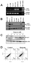
Figure 1
Figure 1. Gal-1 expression analysis
(A). RT-PCR analysis. Five μg of total RNA isolated from each cell line indicated in the figure, was analyzed for the presence of gal-1 transcript by RT-PCR. Equal volumes of these PCR mixtures were separated on 1% agarose gels containing ethidium bromide. GAPDH was amplified as an internal control. (B). Western blot analysis. Equal amount of total cell lysates (20 μg) obtained from the indicated cell lines was separated on 4–20% acrylamide gradient SDS-PAGE gels followed by immunoblotting using anti-gal-1 antibody. Gal-1 and β-actin were identified with arrows. Standard protein markers were loaded in the far right lane. (C). Effect of butyrate on gal-1 expression. LS-180 cells were treated with the indicated concentrations of sodium butyrate prepared in PBS (untreated was indicated with C) for 48 h and analyzed for gal-1 expression in equal amount of cell lysates (20 μg). β-actin was stained to indicate equal protein loading. (D). Analysis of butyrate induced apoptosis. Cells treated with 0 (control) and 5 mM butyrate were subjected to apoptosis assay by flow cytometry as described in the Materials and Methods section. The ordinates show the degree of staining with propidium iodide (FL2-H), indicative of necrosis or late apoptosis, whereas the abscissas show the fluorescence intensity due to annexin V-FITC binding (FL1-H), indicative of apoptosis. The percentages of cells in quadrants, Q1 (necrosis), Q2 (late apoptosis), Q3 (early apoptosis) and Q4 (unstained/live cells) were shown.
Figure 2
Figure 2. Analysis of LGALS1 promoter methylation
(A). MethPrimer analysis of LGALS1 promoter. Promoter region of LGALS1 was analyzed by MethPrimer algorithm as described in Results. The shaded areas represent the CpG clusters. (B). Methylation-specific PCR. Bisulfite-treated genomic DNA from the indicated cell lines was amplified using Methylated- (M) and Unmethylated- (U) specific primers of LGALS1 promoter, as described under Materials and Methods section. The universal unmethylated and methylated human DNA standards were used as control. (C and D). Effect of 5-AzaC on gal-1 expression. LS-180 and Caco-2 cells were treated with 0 (C) and 30 (T) μM 5-AzaC and analyzed by RT-PCR (Panel C) and western blotting (Panel D) for gal-1 expression. GAPDH was used as loading control for RT-PCR. β-Actin was developed to indicate equal protein loading in the western blot.
Figure 3
Figure 3. Gal-1 expression in LS-180 cells. (A). Transient expression of gal-1
LS-180 cells were transfected with vector (C) or gal-1 plasmid (Gal-1) and analyzed by Westernblotting at the indicated time intervals for gal-1 expression. (B). Immunocytochemistry of gal-1 in LS-180 cells. The cells were stained with anti-gal-1 antibody followed by Alexa Fluor-488 coupled secondary antibody. The nuclei were stained with propidium iodide (PI). Images were obtained using Leica Confocal Laser Microscope. The scale bar represents 10 microns. (C). Characterization of cell surface-bound gal-1. Cell surface-bound gal-1 in SW620 (upper panel), and LS-180 (middle and lower panels) cells was analyzed by flow cytometric analysis as described under Materials and Methods section. Fluorescence peaks obtained with preimmune serum (solid line) and anti-gal-1 antibody (dotted line) were shown.
Figure 4
Figure 4. Effects of gal-1 on cellular processes
(A). Cell proliferation assay. Vector and gal-1 plasmid transfected cells were subjected to MTS proliferation assay as described under Materials and Methods. (B). Cell cycle analysis. The transfected cells were subjected to cell cycle analysis by flow cytometry and the G1, S and G2/M phase distribution of cells was quantified using FlowJo software. (C). Wound healing assay. The transiently transfected cells were used in the wound healing assay as described under Materials and Methods. (D). Cell motility assay. The transiently transfected cells were used in motility assay as described under Materials and Methods. * indicates P < 0.05, ** indicates P < 0.01 and *** indicates P < 0.001.
Figure 5
Figure 5. Effects of gal-1 on cell signaling
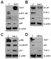
LS-180 cells were transiently transfected with vector (C) and gal-1 plasmid (Gal-1) for 24 h. Cell lysates (20 μg each) were analyzed for: Panel: A. NF-κB signaling molecules - p65, P-p65 and P-IKK α/β; Panel: B. Wnt signaling molecules - β-catenin (β-cat), TCF-1 and TCF-3 and Panel: C. Cell cycle molecules – cyclin D1, phospho-Rb (p-Rb) and p21. (D). Effect of gal-1 knockdown on cell signaling. ATRFLOX cells were transfected either with gal-1 siRNA (siRNA) or control siRNA-A (C) for 48 h and analyzed for the expression of gal-1, p21 and TCF-1. β-actin was stained to indicate equal protein loading.
Figure 6
Figure 6. Analysis of gal-1 induced apoptosis in LS-180 cells
(A). Apoptosis assay. Cells transiently transfected with gal-1 plasmid and vector for up to 48 h and then subjected to apoptosis assay as described in the Materials and Methods section. (B). The effect of CPT on gal-1 induced apoptosis. Cells transfected as above were subjected to apoptosis assay in the presence of 5 μM CPT or DMSO. (C). TMRM assay. Transfected cells were subjected to TMRM assay. Percent change in the TMRM fluorescence in the cells was analyzed using flow cytometry. (D and E). Western blot analysis of different cellular proteins. Lysates obtained from cells transfected with vector (C) or gal-1 plasmid (Gal-1) were subjected to Westernblotting for detecting the indicated proteins. (F). Effect of caspase inhibitors on gal-1-induced apoptosis. Apoptosis assay was carried out on gal-1 plasmid-transfected cells in the presence of DMSO (vehicle control) and 60 and 170 nM caspase-3/7 inhibitor I. ** indicates P < 0.01 and *** indicates P < 0.001.

Similar articles

Cited by

References

    1. Rustgi AK. Molecular genetics and colorectal cancer. Gastroenterology. 1993;104:1223–5. - PubMed
    1. Issa JP. The epigenetics of colorectal cancer. Ann N Y Acad Sci. 2000;910:140–53. discussion 153–5. - PubMed
    1. Shen L, Issa JP. Epigenetics in colorectal cancer. Curr Opin Gastroenterol. 2002;18:68–73. - PubMed
    1. Wheeler JM. Epigenetics, mismatch repair genes and colorectal cancer. Ann R Coll Surg Engl. 2005;87:15–20. - PMC - PubMed
    1. Liu FT, Rabinovich GA. Galectins as modulators of tumour progression. Nat Rev Cancer. 2005;5:29–41. - PubMed

Publication types