

The lymph as a pool of self-antigens
- PMID: 21123113
- PMCID: PMC3052980
- DOI: 10.1016/j.it.2010.10.004
The lymph as a pool of self-antigens
Abstract
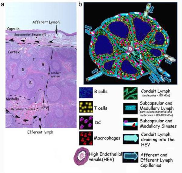
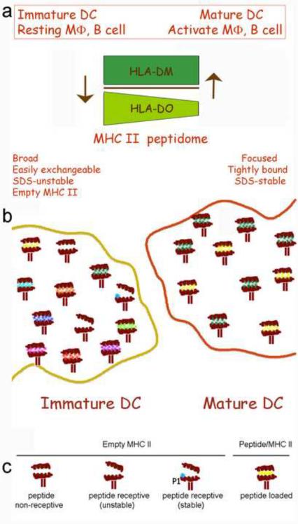
Prenodal lymph is generated from the interstitial fluid that surrounds organs, and thus contains products of organ metabolism and catabolism. New proteomic analyses of lymph have identified proteins and peptides that are derived from capillary extravasation and tissue-specific proteins. Many of these peptides are detected at nanomolar concentrations in the lymph before passage through a regional lymph node. Before entering the node and once inside, proteins and processed peptides are filtered from the lymph by circulating immature dendritic cells (DCs) or non-activated nodal antigen-presenting cells (APCs) (macrophages, B cells and immature DCs). Here, we suggest that this process ensures organ-specific self-antigens are displayed to circulating and nodal APCs, thus contributing to the maintenance of peripheral tolerance.
Copyright © 2010 Elsevier Ltd. All rights reserved.
Figures
Similar articles
-
An expanded self-antigen peptidome is carried by the human lymph as compared to the plasma.PLoS One. 2010 Mar 26;5(3):e9863. doi: 10.1371/journal.pone.0009863. PLoS One. 2010. PMID: 20360855 Free PMC article.
-
Unraveling features of the natural MHC class II peptidome of skin-migrated dendritic cells.Int Immunol. 2012 Jan;24(1):59-69. doi: 10.1093/intimm/dxr096. Epub 2011 Dec 22. Int Immunol. 2012. PMID: 22194283
-
Carrying yourself: self antigen composition of the lymphatic fluid.Lymphat Res Biol. 2013 Sep;11(3):149-54. doi: 10.1089/lrb.2013.0009. Epub 2013 Sep 11. Lymphat Res Biol. 2013. PMID: 24024574 Free PMC article.
-
The Lymphatic Fluid.Int Rev Cell Mol Biol. 2018;337:111-133. doi: 10.1016/bs.ircmb.2017.12.002. Epub 2018 Feb 3. Int Rev Cell Mol Biol. 2018. PMID: 29551158 Review.
-
Exploring the role of antigen presenting cells in male genital tract.Andrologia. 2018 Dec;50(11):e13120. doi: 10.1111/and.13120. Andrologia. 2018. PMID: 30569647 Review.
Cited by
-
Lymphatic system: an active pathway for immune protection.Semin Cell Dev Biol. 2015 Feb;38:83-9. doi: 10.1016/j.semcdb.2014.11.012. Epub 2014 Dec 19. Semin Cell Dev Biol. 2015. PMID: 25534659 Free PMC article. Review.
-
Protein expression profiles of human lymph and plasma mapped by 2D-DIGE and 1D SDS-PAGE coupled with nanoLC-ESI-MS/MS bottom-up proteomics.J Proteomics. 2013 Jan 14;78:172-87. doi: 10.1016/j.jprot.2012.11.013. Epub 2012 Nov 30. J Proteomics. 2013. PMID: 23202415 Free PMC article. Clinical Trial.
-
Promiscuous binding of extracellular peptides to cell surface class I MHC protein.Proc Natl Acad Sci U S A. 2012 Mar 20;109(12):4580-5. doi: 10.1073/pnas.1201586109. Epub 2012 Mar 7. Proc Natl Acad Sci U S A. 2012. PMID: 22403068 Free PMC article.
-
Lymphatic endothelial cells prime naïve CD8+ T cells into memory cells under steady-state conditions.Nat Commun. 2020 Jan 27;11(1):538. doi: 10.1038/s41467-019-14127-9. Nat Commun. 2020. PMID: 31988323 Free PMC article.
-
Steady-state antigen scavenging, cross-presentation, and CD8+ T cell priming: a new role for lymphatic endothelial cells.J Immunol. 2014 Jun 1;192(11):5002-11. doi: 10.4049/jimmunol.1302492. Epub 2014 May 2. J Immunol. 2014. PMID: 24795456 Free PMC article.
References
-
- Heim WJ. On the Chemical Composition of lymph from subcutaneous vessels. Harvard University Press; Cambridge, Mass.: 1932. pp. 553–558.
-
- Yoffey J, et al. Lymphatics, Lymph, and Lymphoid tissue. Harvard University Press; Cambridge, Mass.: 1956.
-
- Levick JR, Michel CC. Microvascular fluid exchange and the revised Starling principle. C. ardiovasc. Res. 2010;87(2):198–210. - PubMed
-
- Aukland K, et al. Protein concentration of lymph and interstitial fluid in the rat tail. Am. J. Physiol. 1984;247(1):4–9. - PubMed
-
- Leak LV, et al. Proteomic analysis of lymph. Proteomics. 2004;4:753–765. - PubMed
MeSH terms
Substances
Grants and funding
LinkOut - more resources
Full Text Sources
Miscellaneous
