. 2011 Feb 11;286(6):4854-70.
doi: 10.1074/jbc.M110.125567. Epub 2010 Dec 1.

Surfactant protein A (SP-A)-mediated clearance of Staphylococcus aureus involves binding of SP-A to the staphylococcal adhesin eap and the macrophage receptors SP-A receptor 210 and scavenger receptor class A

Affiliations

Surfactant protein A (SP-A)-mediated clearance of Staphylococcus aureus involves binding of SP-A to the staphylococcal adhesin eap and the macrophage receptors SP-A receptor 210 and scavenger receptor class A

Zvjezdana Sever-Chroneos et al. J Biol Chem. .

Abstract

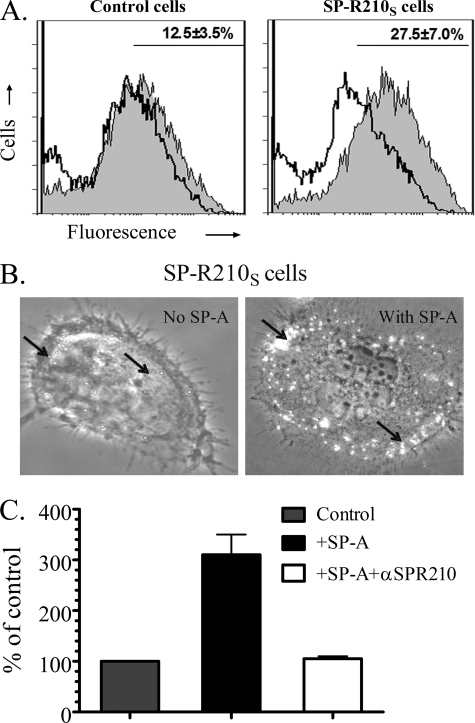
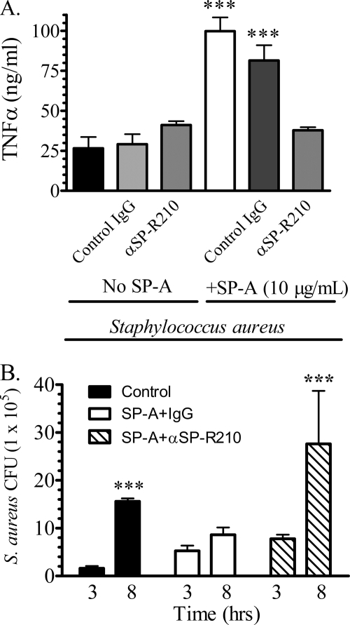
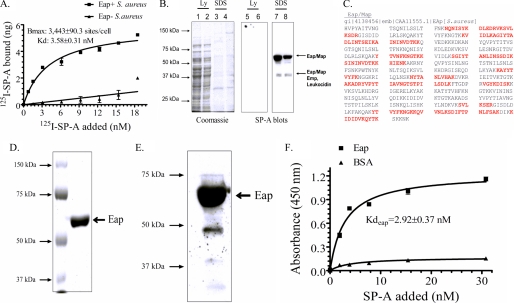
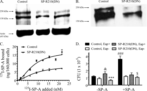
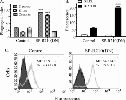
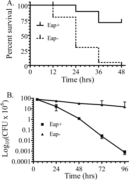
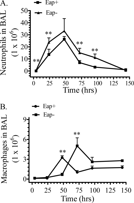
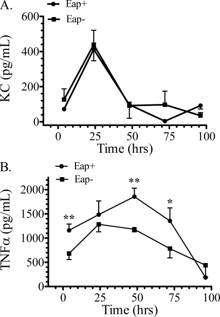
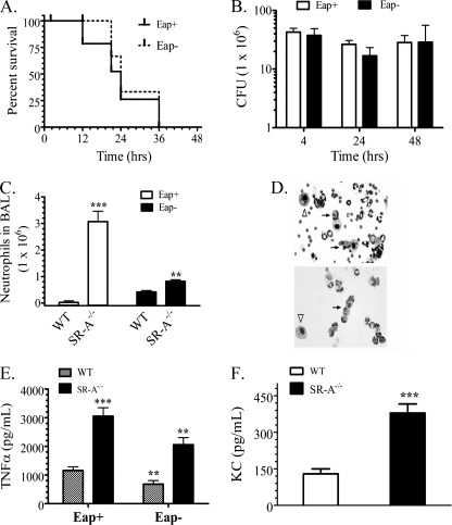
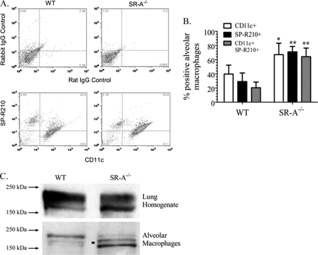
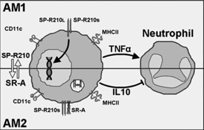
Staphylococcus aureus causes life-threatening pneumonia in hospitals and deadly superinfection during viral influenza. The current study investigated the role of surfactant protein A (SP-A) in opsonization and clearance of S. aureus. Previous studies showed that SP-A mediates phagocytosis via the SP-A receptor 210 (SP-R210). Here, we show that SP-R210 mediates binding and control of SP-A-opsonized S. aureus by macrophages. We determined that SP-A binds S. aureus through the extracellular adhesin Eap. Consequently, SP-A enhanced macrophage uptake of Eap-expressing (Eap(+)) but not Eap-deficient (Eap(-)) S. aureus. In a reciprocal fashion, SP-A failed to enhance uptake of Eap(+) S. aureus in peritoneal Raw264.7 macrophages with a dominant negative mutation (SP-R210(DN)) blocking surface expression of SP-R210. Accordingly, WT mice cleared infection with Eap(+) but succumbed to sublethal infection with Eap- S. aureus. However, SP-R210(DN) cells compensated by increasing non-opsonic phagocytosis of Eap(+) S. aureus via the scavenger receptor scavenger receptor class A (SR-A), while non-opsonic uptake of Eap(-) S. aureus was impaired. Macrophages express two isoforms: SP-R210(L) and SP-R210(S). The results show that WT alveolar macrophages are distinguished by expression of SP-R210(L), whereas SR-A(-/-) alveolar macrophages are deficient in SP-R210(L) expressing only SP-R210(S). Accordingly, SR-A(-/-) mice were highly susceptible to both Eap(+) and Eap(-) S. aureus. The lungs of susceptible mice generated abnormal inflammatory responses that were associated with impaired killing and persistence of S. aureus infection in the lung. In conclusion, alveolar macrophage SP-R210(L) mediates recognition and killing of SP-A-opsonized S. aureus in vivo, coordinating inflammatory responses and resolution of S. aureus pneumonia through interaction with SR-A.


Figures

FIGURE 1.
FIGURE 1.
SP-R210S mediates attachment of SP-A-opsonized S. aureus. A, control and SP-R210S-COS-1 cells were incubated with a 50:1 ratio of FITC-labeled S. aureus alone or after preincubation with 20 μg/ml SP-A. Representative histograms show fluorescence of attached non-opsonized (open histograms) and SP-A-opsonized bacteria (gray histograms). The percentage of cells containing SP-A-opsonized bacteria obtained by gating is shown in the graphs. Data are means ± S.D. (n = 4). B, cell-bound bacteria were visualized by partial bright field illumination of fluorescent bacteria under a ×40 phase-contrast lens. Bacteria (arrows) appear as bright dots on control and SP-R210S-COS-1 cells. C, undifferentiated THP-1 cells were incubated with 1.5 × 10⁶ unopsonized or SP-A-opsonized S. aureus for 1 h at 37 °C in opsonization medium. THP-1 cells were treated with 50 μg/ml control or anti-SP-R210n antibodies for 2 h before infection. Washed cells were then lysed, and cell-associated bacterial cfu were enumerated by serial dilution of lysates on TSB-agar. Data are means ± S.D. (error bars) (n = 3).
FIGURE 2.
FIGURE 2.
SP-R210 mediates TNFα secretion and control of S. aureus growth in macrophages. Mouse bone marrow-derived macrophages were cultured overnight in 24-well dishes at a density of 500,000 cells/well in DMEM, 10% FCS. Serum-deprived macrophages were then preincubated with 20 μg/ml control or anti-SP-R210 antibodies for 1 h before infection with 5 × 10⁶ unopsonized or SP-A-opsonized S. aureus. A, the concentration of TNFα was measured by ELISA in media collected 3 h after infection. Data are means ± S.D. (error bars) (n = 4). ***, p < 0.005 compared with controls in the absence of SP-A. B, infected macrophages were washed in DMEM, and cell-associated bacterial cfu were quantified after serial dilution of cell lysates on tryptic soy broth-agar plates at 3 and 8 h after infection. Data are means ± S.D. (n = 4). ***, p < 0.005 compared with cfu at 3 h after infection.
FIGURE 3.
FIGURE 3.
SP-A binding to S. aureus requires expression of the adhesin Eap. A, binding properties of SP-A to Eap+ or Eap S. aureus Newman were determined by incubating increasing concentrations of 125I-SP-A with 50 × 106 bacteria for 1 h at 37 °C in PBS, 1% BSA without Ca2+. Complexes of bacteria and 125I-SP-A were separated over oil, and bound SP-A was determined using a γ-counter. Data are means ± S.D. (error bars) (n = 4). B, cell wall proteins released from the S. aureus cell wall with either lysostaphin (Ly) (lanes 1, 2, 5, and 6) or 0.4% SDS (lanes 3, 4, 7, and 8) were separated on 10% SDS-polyacrylamide gels. Separated protein was either stained with Coomassie Blue (lanes 1–4) or electrotransferred to nitrocellulose and blotted with biotinylated SP-A (lanes 5–8). MALDI fingerprinting identified the major 64-kDa SP-A-binding protein as the adhesin Eap/Map. The minor 43 kDa band contained peptides from Eap, Emp, and a leukocidin subunit. The results of duplicate experiments are shown. C, the protein sequence of Eap is shown. Identified peptides, shown in red, cover over 50% of the protein. D, native Eap purified from S. aureus Newman (1 μg) was applied on a 10% SDS-polyacrylamide gel and stained with Colloidal Blue. E, purified Eap (1 μg) was visualized by blotting with biotinylated SP-A. F, binding of SP-A to purified Eap was assessed in solid phase assays. Native Eap or BSA adsorbed onto 96-well microtiter plates were incubated with human SP-A (0–20 μg/ml) in blocking buffer for 2 h at 37 °C. Bound SP-A was measured following sequential incubations with rabbit anti-SP-A antibodies and rabbit HRP-conjugated secondary antibody and visualized using 1× tetramethylbenzidine at 450 nm. Data are means ± S.D. (n = 3).
FIGURE 4.
FIGURE 4.
SP-R210 mediates uptake of SP-A-opsonized bacteria. A, dominant negative inhibition of SP-R210 in Raw264.7 macrophages. Western blot analysis assessed expression of SP-R210 in control and SP-R210(DN) macrophages. Proteins (30 μg/lane) were separated on 7% SDS-polyacrylamide gels and electroblotted to nitrocellulose. SP-R210(DN) cell extracts were obtained from two independently derived SP-R210(DN) cells. Probing blots with actin served as loading control. B, control and SP-R210(DN) cells were surface-biotinylated, and extracts were obtained in lysis buffer. Biotinylated protein was precipitated using streptavidin-agarose, and bound protein was separated on 7% SDS-PAGE. Western blot analysis determined the presence of biotinylated SP-R210. A representative blot from two separate experiments is shown. C, binding properties of SP-A to control and SP-R210(DN) cells were measured by incubating cells with increasing concentrations of 125I-SP-A in a 0.1-ml assay volume for 1 h on ice. Bound radioactivity was measured after separation of cells by density centrifugation over oil. Saturation curves were drawn using Prism software. Data shown are means ± S.D. (error bars) (n = 4). D, SP-R210 mediates uptake of SP-A-opsonized Eap+ S. aureus. Control (black and white bars) and SP-R210(DN) (dotted and lined bars) macrophages were infected with a 1:3 ratio of either Eap+ or Eap S. aureus before or after preincubation with SP-A. After washing, macrophage-associated bacterial cfu were quantified by serial dilution of macrophage lysates. Data shown are means ± S.D. (n = 4 triplicate experiments). ###, p < 0.0001 for SP-A opsonized versus unopsonized Eap+ S. aureus in control macrophages (black bars). ***, p < 0.001 for Eap- versus Eap+ S. aureus infection of SP-R210(DN) macrophages in the absence of SP-A. &, p < 0.05 for Eap+ S. aureus infection between control and SP-R210(DN) cells in the absence of SP-A. $, p < 0.05 for SP-R210(DN) macrophages infected with Eap S. aureus in the presence versus absence of SP-A.
FIGURE 5.
FIGURE 5.
Increased expression and function of SR-A in SP-R210(DN) macrophages. A, phagocytic activity of control and SP-R210(DN) macrophages was assessed using FITC-labeled S. aureus, E. coli, or yeast zymosan bioparticles. Control and SP-R210(DN) were incubated with a 10:1 ratio of the indicated bioparticles for 30 min at 37 °C. Data are expressed as phagocytic index, representing the percentage of cells containing internalized bioparticles. Data are means ± S.D. (error bars) (n = 12). ***, p < 0.001 compared with control macrophages. B, scavenger receptor activity of control and SP-R210(DN) was assessed using fluorescently labeled acetylated LDL (DiAcLDL) as an endocytic ligand of scavenger receptors. Native fluorescent LDL (DiLDL) was used as control. Macrophages were incubated with 0.8 μg/ml native fluorescent LDL or fluorescently labeled acetylated LDL for 30 min at 37 °C. Uptake of LDLs was analyzed by flow cytometry. Data are means ± S.D. (n = 8). ***, p < 0.001 compared with control macrophages. C, surface expression of the scavenger receptor SR-A in control and SP-R210(DN) cells was assessed using a monoclonal rat anti-mouse SR-A antibody clone 2F8. Grey-shaded and unshaded histograms show staining with isotype control and anti-SR-A antibodies, respectively. Linear gating was used to calculate fluorescent intensity, and the percentage of SR-A-positive cells in control and SP-R210(DN) cell cultures is shown in histogram insets. Data are means ± S.D. (n = 8).
FIGURE 6.
FIGURE 6.
Survival and pulmonary bacterial clearance of mice infected with Eap+ or EapS. aureus. A, survival of 6–8-week-old C57BL/6 male mice was monitored at 12-h intervals after intranasal infection with 300 × 106 cfu of Eap+ or Eap S. aureus Newman. Data shown are from a total of n = 40 mice/group in three independent experiments. Survival curves were generated using the Kaplan-Meier method. The mean survival of mice infected with Eap S. aureus was 24 h. Differences in survival were statistically significant with p < 0.0001. Statistical analysis of survival curves was performed using the Gehan-Breslow-Wilcoxon test tool using Prism software. B, pulmonary clearance of intranasal infection of mice with 200 × 106 cfu of Eap+ and Eap S. aureus was monitored in lung homogenates at the indicated time intervals. Data shown are means ± S.E. (error bars) (n = 7–9 at the 4, 24, and 72 h time points, n = 4 at the 96 h time point).
FIGURE 7.
FIGURE 7.
Cytospin analysis of alveolar lavage from mice infected with EAP+ and EapS. aureus. Bronchoalveolar lavage was collected at 4 h (A and B), 24 h (C and D), 48 h (E and F), 72 h (G and H), and 96 h (I and J) after intranasal infection of mice with 200 × 106 cfu Eap+ (A, C, E, G, and I, left panels) or Eap (B, D, F, H, and J, right panels) S. aureus. Cells were deposited onto glass slides by cytospin centrifugation and stained with HEMA-3 to evaluate inflammatory cells and cell-associated bacteria. Images in E and F were captured at ×40 magnification. All other images were photographed at ×20 magnification. Open and closed head arrows (A–F, H, and J) point to infected neutrophils and macrophages, respectively. The diamond head arrows (J) indicate vacuolated cells.
FIGURE 8.
FIGURE 8.
Recruitment of neutrophils and macrophages in mice infected with Eap+ and EapS. aureus. Cellular infiltrates were evaluated in bronchoalveolar lavage (BAL) after intranasal infection of WT mice with 200 × 106 cfu Eap+ or Eap S. aureus. Total cell numbers were counted using a hemacytometer. Cell types were identified by differential staining following cytospin centrifugation of lavaged cells. Counting of neutrophils (A) and macrophages (B) in 5–10 microscopic fields determined the percentage of each cell type in bronchoalveolar lavage. The percentage of each cell type was multiplied by the total number of cells in bronchoalveolar lavage to obtain the number of neutrophils and macrophages. Data shown are means ± S.E. (n = 7–9 mice at the 4, 24, and 72 h time points; n = 4–6 mice at the 96 h time point). **, p < 0.03 indicates significant differences in neutrophil and macrophage numbers between Eap+ and Eap S. aureus-infected mice at the indicated time points.
FIGURE 9.
FIGURE 9.
Levels of KC and TNFα in WT mice infected with Eap+ or EapS. aureus. The concentration of KC (A) and TNFα (B) was determined by ELISA in lung homogenates after infection of mice with 200 × 106 cfu Eap+ or Eap S. aureus. Data shown are means ± S.E. (n = 7–9 mice at the 4, 24, and 72 h time points; n = 4–6 mice at the 96 h time point). Statistical differences in TNFα levels at each time point are p < 0.01 (**) at the 4 and 48 h time points and p < 0.05 (*) at the 72 h time point.
FIGURE 10.
FIGURE 10.
Assessment of pneumonia in SR-A−/− mice infected with Eap+ and EapS. aureus. A, survival of 6–8-week-old SR-A−/− mice was monitored at 12-h intervals after infection with 300 × 106 cfu of Eap+ or Eap S. aureus Newman. Data shown are from a total of n = 20 mice/group from two independent experiments. Survival curves were generated using the Kaplan-Meier method. B, pulmonary clearance after infection with 200 × 106 cfu of Eap+ or Eap S. aureus was monitored in lung homogenates at the indicated time intervals. Data shown are mean ± S.E. (error bars) (n = 6–8 at the 4, 24, and 48 h time points). C, the number of neutrophils in lavage 4 h after infection was counted as described in the legend to Fig. 8. Data shown are means ± S.E. (error bars) (n = 8) in two independent experiments. D, cytospin analysis of lung lavage 4 h after infection with the indicated S. aureus strains. E, the concentration of TNFα was measured in lung homogenates 4 h after infection with the indicated S. aureus strains. Data shown are mean ± S.E. (n = 8) in two independent experiments per group. F, the concentration of KC was measured in lung homogenates 4 h after infection with Eap+ S. aureus. Data are mean ± S.E. (n = 8) in two independent experiments.
FIGURE 11.
FIGURE 11.
Differential expression of SP-R210 isoforms in WT and SR-A−/− mice. Flow cytometric analysis determined expression of CD11c and SP-R210 on alveolar macrophages from WT and SR-A−/− mice. A, representative dual color histograms following staining of alveolar macrophages with either isotype-matched IgG in upper panels or anti-CD11c and anti-SP-R210 antibodies in lower panels. B, combined flow cytometric data were expressed as a percentage of single or double positive cells for the indicated markers in WT or SR-A−/− alveolar macrophages. Data are means ± S.E. (error bars) (n = 6 independent experiments on pooled alveolar macrophages from 5 mice/genotype). *, p < 0.05; **, p < 0.01 SR-A−/− compared with WT mice. C, lung or alveolar macrophage extracts were prepared in lysis buffer, separated on 7% SDS-PAGE, and evaluated by Western blot analysis using anti-SP-R210 antibodies. Lanes were loaded with 20 μg of protein. Blots are representative of two separate experiments.
FIGURE 12.
FIGURE 12.
Proposed interaction of SP-R210 and SR-A in alveolar macrophages. Resting alveolar macrophages AM1 express SP-R210L. Ligation of SP-R210L with SP-A-opsonized bacteria in AM1 macrophages induces macrophage activation and secretion of TNFα, timing recruitment and activation of neutrophils. Polarization of AM1 to AM2 macrophages results in expression of SP-R210S and SR-A, coordinating phagocytosis via an SP-R210S·SR-A complex with regulation of the inflammatory response.

Similar articles

Cited by

References

    1. Defres S., Marwick C., Nathwani D. (2009) Eur. Respir. J. 34, 1470–1476 - PubMed
    1. Murray R. J., Robinson J. O., White J. N., Hughes F., Coombs G. W., Pearson J. C., Tan H. L., Chidlow G., Williams S., Christiansen K. J., Smith D. W. (2010) PLoS One 5, e8705 - PMC - PubMed
    1. Sinha B., Fraunholz M. (2010) Int. J. Med. Microbiol. 300, 170–175 - PubMed
    1. Foster T. J. (2009) Vet. Dermatol. 20, 456–470 - PubMed
    1. Floros J., Wang G., Mikerov A. N. (2009) Crit. Rev. Eukaryot. Gene Expr. 19, 125–137 - PMC - PubMed

Publication types

MeSH terms

Substances