

Induction of caspase activation and cleavage of the viral nucleocapsid protein in different cell types during Crimean-Congo hemorrhagic fever virus infection
- PMID: 21123175
- PMCID: PMC3030327
- DOI: 10.1074/jbc.M110.149369
Induction of caspase activation and cleavage of the viral nucleocapsid protein in different cell types during Crimean-Congo hemorrhagic fever virus infection
Abstract
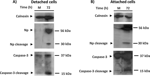
Regulation of apoptosis during infection has been observed for several viral pathogens. Programmed cell death and regulation of apoptosis in response to a viral infection are important factors for host or virus survival. It is not known whether Crimean-Congo hemorrhagic fever virus (CCHFV) infection regulates the apoptosis process in vitro. This study for the first time suggests that CCHFV induces apoptosis, which may be dependent on caspase-3 activation. This study also shows that the coding sequence of the S segment of CCHFV contains a proteolytic cleavage site, DEVD, which is conserved in all CCHFV strains. By using different recombinant expression systems and site-directed mutagenesis, we demonstrated that this motif is subject to caspase cleavage. We also demonstrate that CCHFV nucleocapsid protein (NP) is cleaved into a 30-kDa fragment at the same time as caspase activity is induced during infection. Using caspase inhibitors and cells lacking caspase-3, we clearly demonstrate that the cleavage of NP is caspase-3-dependent. We also show that the inhibition of apoptosis induced progeny viral titers of ∼80-90%. Thus, caspase-3-dependent cleavage of NP may represent a host defense mechanism against lytic CCHFV infection. Taken together, these data suggest that the most abundant protein of CCHFV, which has several essential functions such as protection of viral RNA and participation in various processes in the replication cycle, can be subjected to cleavage by host cell caspases.
Figures
Similar articles
-
Recent advances in understanding Crimean-Congo hemorrhagic fever virus.F1000Res. 2018 Oct 29;7:F1000 Faculty Rev-1715. doi: 10.12688/f1000research.16189.1. eCollection 2018. F1000Res. 2018. PMID: 30416710 Free PMC article. Review.
-
Heat Shock Protein 70 Family Members Interact with Crimean-Congo Hemorrhagic Fever Virus and Hazara Virus Nucleocapsid Proteins and Perform a Functional Role in the Nairovirus Replication Cycle.J Virol. 2016 Sep 29;90(20):9305-16. doi: 10.1128/JVI.00661-16. Print 2016 Oct 15. J Virol. 2016. PMID: 27512070 Free PMC article.
-
Structure of Crimean-Congo hemorrhagic fever virus nucleoprotein: superhelical homo-oligomers and the role of caspase-3 cleavage.J Virol. 2012 Nov;86(22):12294-303. doi: 10.1128/JVI.01627-12. Epub 2012 Sep 5. J Virol. 2012. PMID: 22951837 Free PMC article.
-
Rescue of Infectious Recombinant Hazara Nairovirus from cDNA Reveals the Nucleocapsid Protein DQVD Caspase Cleavage Motif Performs an Essential Role other than Cleavage.J Virol. 2019 Jul 17;93(15):e00616-19. doi: 10.1128/JVI.00616-19. Print 2019 Aug 1. J Virol. 2019. PMID: 31118258 Free PMC article.
-
The Role of Nucleocapsid Protein (NP) in the Immunology of Crimean-Congo Hemorrhagic Fever Virus (CCHFV).Viruses. 2024 Sep 30;16(10):1547. doi: 10.3390/v16101547. Viruses. 2024. PMID: 39459881 Free PMC article. Review.
Cited by
-
Recent advances in understanding Crimean-Congo hemorrhagic fever virus.F1000Res. 2018 Oct 29;7:F1000 Faculty Rev-1715. doi: 10.12688/f1000research.16189.1. eCollection 2018. F1000Res. 2018. PMID: 30416710 Free PMC article. Review.
-
Fine epitope mapping of the central immunodominant region of nucleoprotein from Crimean-Congo hemorrhagic fever virus (CCHFV).PLoS One. 2014 Nov 3;9(11):e108419. doi: 10.1371/journal.pone.0108419. eCollection 2014. PLoS One. 2014. PMID: 25365026 Free PMC article.
-
Comparative characterization of Crimean-Congo hemorrhagic fever virus cell culture systems with application to propagation and titration methods.Virol J. 2023 Jun 19;20(1):128. doi: 10.1186/s12985-023-02089-w. Virol J. 2023. PMID: 37337294 Free PMC article.
-
Virus-Derived DNA Forms Mediate the Persistent Infection of Tick Cells by Hazara Virus and Crimean-Congo Hemorrhagic Fever Virus.J Virol. 2021 Nov 23;95(24):e0163821. doi: 10.1128/JVI.01638-21. Epub 2021 Oct 6. J Virol. 2021. PMID: 34613808 Free PMC article.
-
Cleavage of the Junin virus nucleoprotein serves a decoy function to inhibit the induction of apoptosis during infection.J Virol. 2013 Jan;87(1):224-33. doi: 10.1128/JVI.01929-12. Epub 2012 Oct 17. J Virol. 2013. PMID: 23077297 Free PMC article.
References
-
- Zimmermann K. C., Bonzon C., Green D. R. (2001) Pharmacol. Ther. 92, 57–70 - PubMed
-
- Thornberry N. A. (1998) Chem. Biol. 5, R97–R103 - PubMed
-
- Thornberry N. A., Lazebnik Y. (1998) Science 281, 1312–1316 - PubMed
-
- Tarodi B., Subramanian T., Chinnadurai G. (1994) Virology 201, 404–407 - PubMed
-
- Gadaleta P., Vacotto M., Coulombié F. (2002) Virus Res. 86, 87–92 - PubMed
Publication types
MeSH terms
Substances
LinkOut - more resources
Full Text Sources
Research Materials
Miscellaneous
