

Pulmonary thin-section CT findings in acute Moraxella catarrhalis pulmonary infection
- PMID: 21123308
- PMCID: PMC3473838
- DOI: 10.1259/bjr/42762966
Pulmonary thin-section CT findings in acute Moraxella catarrhalis pulmonary infection
Abstract
Objective: Moraxella catarrhalis is an important pathogen in the exacerbation of chronic obstructive pulmonary disease. The aim of this study was to assess the clinical and pulmonary thin-section CT findings in patients with acute M. catarrhalis pulmonary infection.
Methods: Thin-section CT scans obtained between January 2004 and March 2009 from 292 patients with acute M. catarrhalis pulmonary infection were retrospectively evaluated. Clinical and pulmonary CT findings in the patients were assessed. Patients with concurrent infection including Streptococcus pneumoniae (n = 72), Haemophilus influenzae (n = 61) or multiple pathogens were excluded from this study.
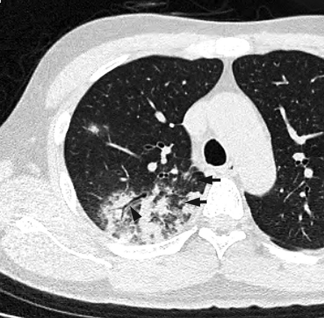
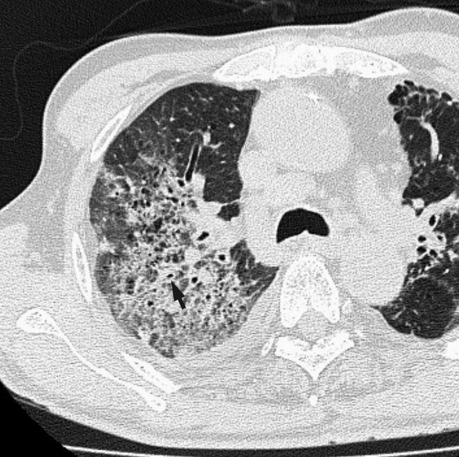
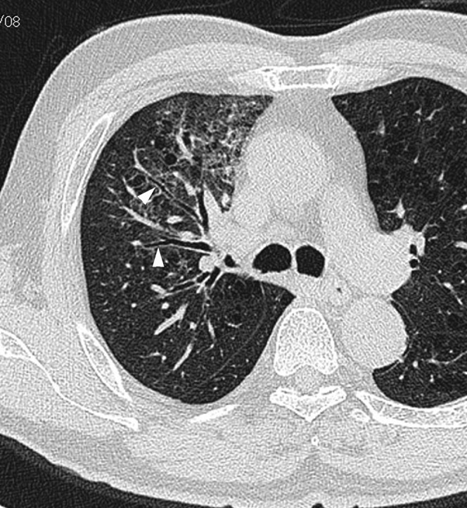
Results: The study group comprised 109 patients (66 male, 43 female; age range 28-102 years; mean age 74.9 years). Among the 109 patients, 34 had community-acquired and 75 had nosocomial infections. Underlying diseases included pulmonary emphysema (n = 74), cardiovascular disease (n = 44) or malignant disease (n = 41). Abnormal findings were seen on CT scans in all patients and included ground-glass opacity (n = 99), bronchial wall thickening (n = 85) and centrilobular nodules (n = 79). These abnormalities were predominantly seen in the peripheral lung parenchyma (n = 99). Pleural effusion was found in eight patients. No patients had mediastinal and/or hilar lymph node enlargement.
Conclusions: M. catarrhalis pulmonary infection was observed in elderly patients, often in combination with pulmonary emphysema. CT manifestations of infection were mainly ground-glass opacity, bronchial wall thickening and centilobular nodules.
Figures

Similar articles
-
Thin-section CT findings in Pseudomonas aeruginosa pulmonary infection.Br J Radiol. 2012 Dec;85(1020):1533-8. doi: 10.1259/bjr/54468236. Epub 2012 Jul 27. Br J Radiol. 2012. PMID: 22844034 Free PMC article.
-
Radiological findings in acute Haemophilus influenzae pulmonary infection.Br J Radiol. 2012 Feb;85(1010):121-6. doi: 10.1259/bjr/48077494. Epub 2011 Jan 11. Br J Radiol. 2012. PMID: 21224303 Free PMC article.
-
Bacteriological incidence in pneumonia patients with pulmonary emphysema: a bacterial floral analysis using the 16S ribosomal RNA gene in bronchoalveolar lavage fluid.Int J Chron Obstruct Pulmon Dis. 2017 Jul 20;12:2111-2120. doi: 10.2147/COPD.S140901. eCollection 2017. Int J Chron Obstruct Pulmon Dis. 2017. PMID: 28790814 Free PMC article.
-
Moraxella catarrhalis bacteraemia associated with prosthetic vascular graft infection.J Med Microbiol. 2010 Feb;59(Pt 2):245-250. doi: 10.1099/jmm.0.013789-0. Epub 2009 Oct 22. J Med Microbiol. 2010. PMID: 19850707 Review.
-
Potential impact of a Moraxella catarrhalis vaccine in COPD.Vaccine. 2019 Sep 3;37(37):5551-5558. doi: 10.1016/j.vaccine.2016.12.066. Epub 2017 Feb 6. Vaccine. 2019. PMID: 28185742 Free PMC article. Review.
Cited by
-
Clinical characteristics of community-acquired pneumonia due to Moraxella catarrhalis in adults: a retrospective single-centre study.BMC Infect Dis. 2020 Nov 10;20(1):821. doi: 10.1186/s12879-020-05564-9. BMC Infect Dis. 2020. PMID: 33172398 Free PMC article.
-
Thin-section CT findings in Pseudomonas aeruginosa pulmonary infection.Br J Radiol. 2012 Dec;85(1020):1533-8. doi: 10.1259/bjr/54468236. Epub 2012 Jul 27. Br J Radiol. 2012. PMID: 22844034 Free PMC article.
-
Differential diagnosis of pulmonary infections in immunocompromised patients using high-resolution computed tomography.Eur Radiol. 2019 Nov;29(11):6089-6099. doi: 10.1007/s00330-019-06235-3. Epub 2019 May 6. Eur Radiol. 2019. PMID: 31062135
-
Differentiation of pulmonary complications with extensive ground-glass attenuation on high-resolution CT in immunocompromised patients.Jpn J Radiol. 2021 Sep;39(9):868-876. doi: 10.1007/s11604-021-01122-8. Epub 2021 May 4. Jpn J Radiol. 2021. PMID: 33945100 Free PMC article.
-
Exploring the Application of Metagenomic Next-Generation Sequencing in the Diagnosis of Unexplained Pulmonary Infection.Int J Gen Med. 2024 May 27;17:2465-2474. doi: 10.2147/IJGM.S459373. eCollection 2024. Int J Gen Med. 2024. PMID: 38826507 Free PMC article.
References
-
- Frosch P, Kolle W. Die Mikrokokken Flugge C, ed. Die Mikroorganismen. Leipzig: Verlag von Vogel, 1956;154–5
-
- Wright PW, Wallace RJ, Shepherd JR. A descriptive study of 42 cases of Branhamella catarrhalis pneumonia. Am J Med 1990;88:2S–8S - PubMed
-
- West M, Berk SL, Smith JK. Branhamella catarrhalis pneumonia. Southern Med J 1982;75:1021–3 - PubMed
-
- Wallace RJ, Musher DM. In honor of Dr. Sara Braham, a star is born. The realization of Brahamella catarrhalis as a respiratory pathogen. Chest 1986;90:447–50 - PubMed
-
- Kovatch AL, Wald ER, Michaels RH. Beta-lactamase producing Brahamella catarrhalis causing otitis media in children. J Pediatrics 1983;102:261–4 - PubMed
