. 2011 Feb;85(4):1484-94.
doi: 10.1128/JVI.02167-10. Epub 2010 Dec 1.

Crucial role for prion protein membrane anchoring in the neuroinvasion and neural spread of prion infection

Affiliations

Crucial role for prion protein membrane anchoring in the neuroinvasion and neural spread of prion infection

Mikael Klingeborn et al. J Virol. 2011 Feb.

Abstract

In nature prion diseases are usually transmitted by extracerebral prion infection, but clinical disease results only after invasion of the central nervous system (CNS). Prion protein (PrP), a host-encoded glycosylphosphatidylinositol (GPI)-anchored membrane glycoprotein, is necessary for prion infection and disease. Here, we investigated the role of the anchoring of PrP on prion neuroinvasion by studying various inoculation routes in mice expressing either anchored or anchorless PrP. In control mice with anchored PrP, intracerebral or sciatic nerve inoculation resulted in rapid CNS neuroinvasion and clinical disease (154 to 156 days), and after tongue, ocular, intravenous, or intraperitoneal inoculation, CNS neuroinvasion was only slightly slower (193 to 231 days). In contrast, in anchorless PrP mice, these routes resulted in slow and infrequent CNS neuroinvasion. Only intracerebral inoculation caused brain PrPres, a protease-resistant isoform of PrP, and disease in both types of mice. Thus, anchored PrP was an essential component for the rapid neural spread and CNS neuroinvasion of prion infection.


Figures
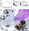
FIG. 1.
FIG. 1.
PrPres deposition in sciatic nerve and CNS following intracerebral and sciatic nerve scrapie inoculation of tg44+/+ mice. (A) Relative levels of PrPres deposition detected by immunoblotting in brain of tg44+/+ mice after scrapie inoculation via intracerebral (i.c.), intraneural (i.n.), and perineural (p.n.) routes. Solid symbols represent mice with clinical neurological signs as previously described (17). Relative amount of brain PrPres detected by immunoblotting is defined in Materials and Methods. (B) Immunoblot of PrPres in sciatic nerve and CNS following i.c. (lanes 1 to 4) and i.n. (lanes 5 to 10) scrapie inoculation. Brain samples from clinical C57BL/10 mice (WT) inoculated via indicated routes are in lanes 1 and 5. Tissues from an i.c. inoculated tg44+/+ mouse are in lanes 2 to 4 (308 dpi). Brain and/or lumbar spinal cord samples from two i.n. inoculated tg44+/+ mice are in lanes 6 and 7 (600 dpi) and lane 8 (600 dpi). Ipsilateral sciatic nerves are shown for two i.n. inoculated tg44+/+ mice in lanes 9 and 10 (both 600 dpi). Lane 8 was overexposed to show PrPres in lumbar spinal cord. Lanes 1, 2, and 5 were loaded with 0.25 mg, and all other lanes were loaded with a 1-mg tissue equivalent. Approximate molecular sizes of 18 and 22 kDa are indicated. (C to F) Immunohistochemical detection of PrPres deposits in sciatic nerve and lumbar spinal cord following sciatic nerve scrapie inoculation in tg44+/+ mice. (C) Extensive PrPres deposition was found in the ipsilateral sciatic nerve at 454 dpi. (D) Numerous amyloid plaques (arrows) seen by hematoxylin and eosin staining of the sciatic nerve seen in panel C. (E) PrPres deposition was found in nerve roots (N) and white matter tracts (WM) but only rarely in gray matter tracts (GM) on the ventral aspect of the lumbar spinal cord at 600 dpi. (F) The boxed area in panel E magnified. Arrows show blood vessels surrounded by PrPres. Bars, 100 μm (C, D, and E) and 50 μm (F).
FIG. 2.
FIG. 2.
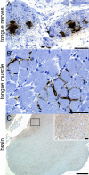
(A) Immunohistochemical detection of PrPres deposits in tongue nerves at 600 days following scrapie inoculation in the tongue of a tg44+/+ mouse. Arrows indicate PrPres deposits in close association with neurolemma. (B) Widespread PrPres accumulation mostly along capillaries between muscle cells in tongue of mouse from panel A. (C) Sagittal section of medullar area of brain stem of the same mouse showing no detectable PrPres. Cerebellum and fourth ventricle are visible in the upper left corner. Inset shows higher magnification of boxed area. Light brown diffuse staining is PrPsen, which is also seen in uninoculated animals (data not shown), and is easily distinguished from PrPres (panels A and B) in tg44+/+ mice. Bars, 50 μm (A, B, and inset in C) and 500 μm (C).
FIG. 3.
FIG. 3.
PrPres deposition in brain and eyes following ocular scrapie inoculation in tg44+/+ mice. (A) Relative amount of PrPres deposition detected in brain by immunoblotting at various times after inoculation. Solid symbols represent mice with clinical neurological signs. (B) Immunoblot of PrPres in brain and eyes following ocular scrapie inoculation. High levels of PrPres can be seen at terminal stage of disease in brains (Br) of an i.o. inoculated C57BL/10 (WT) mouse (lane 1), an i.c. inoculated tg44+/+ mouse at 308 dpi (lane 2), and an i.o. inoculated tg44+/+ mouse at 469 dpi (lane 3), whereas no PrPres was detected in a 520-day-old uninoculated (un) tg44+/+ mouse (lane 4). In lanes 5 to 10, brains and eyes from two other i.o. inoculated tg44+/+ mice are shown (animal 286, 441 dpi; animal 291, 435 dpi). Ipsilateral (I) eyes (lanes 7 and 10) have high levels of PrPres, while the contralateral (C) eyes (lanes 6 and 9) have undetectable levels of PrPres. Brains had either moderate (lane 8) or low (lane 5) levels of PrPres. Lanes 1 to 3 were loaded with 0.25 mg, and all other lanes were loaded with a 1-mg tissue equivalent. Approximate molecular sizes of 18 and 22 kDa are indicated by the two bars on the right hand side. (C) Immunohistochemical staining using monoclonal antibody D13 shows PrPres amyloid plaque deposition in a sagittal section of the cerebellum of a tg44+/+ mouse at 357 days after ocular scrapie inoculation. (D) Higher magnification of panel C shows perivascular PrPres plaques in the molecular layers (M), as well as in meninges between two lobules, around blood vessels (arrows) and in the granular layer (G) of the third cerebellar lobule. Bars, 500 μm (C) and 100 μm (D).
FIG. 4.
FIG. 4.
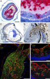
Immunohistochemical and immunofluorescence detection of PrPres deposition in eye and optic nerve at 232 dpi after ocular scrapie inoculation in several tg44+/+ mice. (A) Extensive PrPres deposition (red, Fast Red staining) in retinal ganglion layer at 232 dpi. The eye was cut at an angle tangential to the ganglion layer, resulting in the appearance of a flap of ganglion layer extending into the vitreal space in the center of the section. Fewer PrPres deposits are present in the inner nuclear layer (INL) and only rarely were deposits detected in the outer nuclear layer (ONL) or other retinal cell layers outside INL. (B) At higher magnification of the boxed area in panel A, PrPres deposits are evident in the ganglion layer, and many PrPres plaques have a distinct perivascular appearance (arrows). (C) In a different mouse at 232 dpi the section shows widespread PrPres accumulation (brown, DAB staining) in the optic nerve entering the retina as well as in the ganglion layer of the retina and in the vitreous fluid. (D) At higher magnification of the boxed area in panel C, PrPres can be seen in optic nerve (ON), vitreous fluid (VF), ganglion layer (G), and inner nuclear layer (INL) and around two retinal blood vessels (arrows). PrPres plaques adjacent to the pia mater of optic nerve are indicated by arrowheads. (E) Double immunofluorescence staining of the optic nerve of mouse shown in panels C and D at the level of entry into the retina. PrPres deposits (red) and an extensive network of GFAP-expressing astrocytes (green) can be seen in the optic nerve. This astrocyte network was also seen in optic nerve from uninfected tg44+/+ mice (data not shown). Arrows indicate PrPres plaques in close proximity to the pia mater on the edge of the nerve. (F) Double immunofluorescence staining of retina at high magnification shows amyloid PrPres deposits (red), mostly perivascular and in close proximity to perivascular GFAP-expressing astrocytes (green). Close to the lower edge of the picture, the outer nuclear layer of the retina stains strongly with the nuclear stain DAPI (blue). (G) Iba1-positive microglia or macrophages (green) were detected in close proximity to PrPres deposits (red) in the retinal ganglion layer. Most of the Iba1-positive cells have large plump cell bodies (arrows) and thickened processes, suggesting an activated phenotype. Bars, 500 μm (A and C), 100 μm (D and E), and 50 μm (B, F, and G).
FIG. 5.
FIG. 5.
(A) Relative amount of PrPres deposition detected by immunoblotting in brain of tg44+/+ mice after intravenous (i.v.) and intraperitoneal (i.p.) scrapie inoculation. Solid symbols represent mice with clinical neurological signs. (B) Immunoblot of PrPres in brain and after i.v. or i.p. scrapie inoculation. Brains from three C57BL/10 (WT) mice (lane 1, i.c. at 144 dpi; lane 2, i.v. at 197 dpi; lane 3, i.p. at 188 dpi) and a tg44+/+ mouse (lane 4, i.c. at 308 dpi) at terminal stage of disease are shown. Brain from i.v. inoculated tg44+/+ mice at 603 dpi (lane 5), 560 dpi (lane 6), and 483 dpi (lane 7) showed various levels of PrPres. Brains of i.p. inoculated tg44+/+ mice showed undetectable PrPres (lane 8, 601 dpi) or very low PrPres (lanes 9 and 10, 601 and 580 dpi, respectively). Lanes 5 and 6 and 8 to 10 were loaded with 1.0 mg, and all other lanes were loaded with 0.25-mg tissue equivalents. Approximate molecular masses of 18 and 22 kDa are indicated on the right-hand side. (C and D) Immunohistochemical staining using monoclonal antibody D13 shows PrPres amyloid plaque deposition in brain after intraperitoneal scrapie inoculation of tg44+/+ mice. (C) PrPres plaques (red, Fast Red) were detected in the choroid plexus (arrow) and radiating out from blood vessels (arrowheads) in the meninges of the 10th cerebellar lobule and the fissure between the 9th and 10th cerebellar lobules at 740 days after i.p. inoculation. (D) In another mouse at 601 dpi after i.p. inoculation, PrPres deposits (brown, DAB stain) were found only along the meninges in the posterior portion of the cerebral cortex. Bar, 200 μm.
FIG. 6.
FIG. 6.
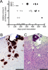
The effect of blood-brain barrier manipulation on PrPres deposition in brain of tg44+/+ mice after intravenous scrapie inoculation. (A) Relative amount of PrPres detected by immunoblotting in brains of tg44+/+ mice after intravenous scrapie inoculation followed by a mock intracerebral needle stab (i.v. + stab) was higher and detected earlier than after intravenous inoculation alone. Solid symbols represent mice with clinical neurological signs, and open symbols represent mice without clinical signs. (B) Immunohistochemical staining with monoclonal antibody D13 at the site of the mock needle stab in the cerebral cortex at 225 dpi after i.v.+stab inoculation in a tg44+/+ mouse. A large PrPres plaque is evident at the dorsal surface adjacent to the pia mater. (C) H&E staining shows disruption of several neuronal cell layers along the needle track corresponding to locations of PrPres amyloid plaques seen in panel B. Bar, 200 μm.

References

    1. Atherton, S. S., C. K. Newell, M. Y. Kanter, and S. W. Cousins. 1991. Retinitis in euthymic mice following inoculation of murine cytomegalovirus (MCMV) via the supraciliary route. Curr. Eye Res. 10:667-677. - PubMed
    1. Ayers, J. I., A. E. Kincaid, and J. C. Bartz. 2009. Prion strain targeting independent of strain-specific neuronal tropism. J. Virol. 83:81-87. - PMC - PubMed
    1. Baldauf, E., M. Beekes, and H. Diringer. 1997. Evidence for an alternative direct route of access for the scrapie agent to the brain bypassing the spinal cord. J. Gen. Virol. 78:1187-1197. - PubMed
    1. Bartz, J. C., C. Dejoia, T. Tucker, A. E. Kincaid, and R. A. Bessen. 2005. Extraneural prion neuroinvasion without lymphoreticular system infection. J. Virol. 79:11858-11863. - PMC - PubMed
    1. Bartz, J. C., A. E. Kincaid, and R. A. Bessen. 2003. Rapid prion neuroinvasion following tongue infection. J. Virol. 77:583-591. - PMC - PubMed

Publication types

LinkOut - more resources