. 2011 Feb 1;71(3):842-51.
doi: 10.1158/0008-5472.CAN-10-1066. Epub 2010 Dec 1.

BCR/ABL stimulates WRN to promote survival and genomic instability

Affiliations

BCR/ABL stimulates WRN to promote survival and genomic instability

Artur Slupianek et al. Cancer Res. .

Abstract

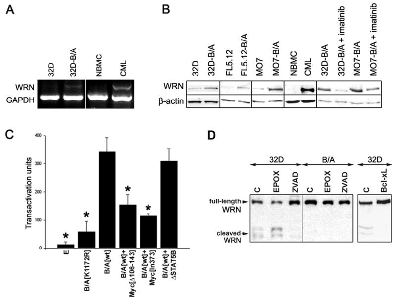
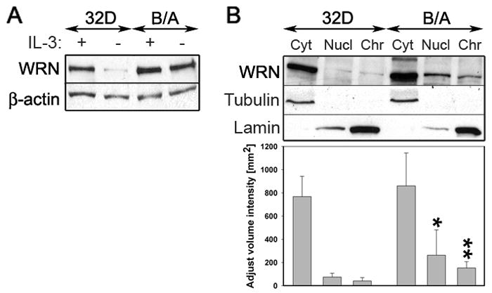
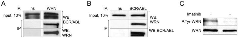
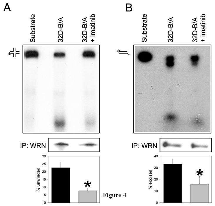
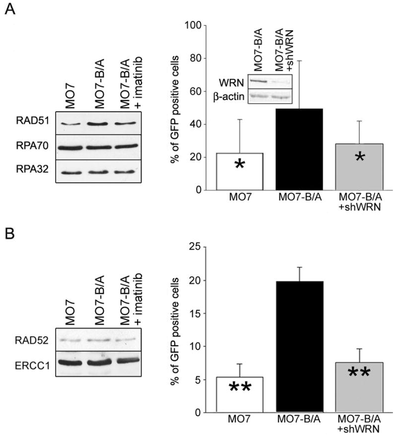
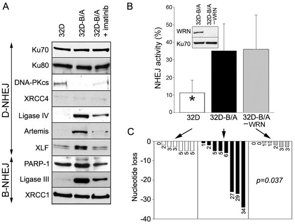
BCR/ABL-transformed chronic myeloid leukemia (CML) cells accumulate numerous DNA double-strand breaks (DSB) induced by reactive oxygen species (ROS) and genotoxic agents. To repair these lesions BCR/ABL stimulate unfaithful DSB repair pathways, homologous recombination repair (HRR), nonhomologous end-joining (NHEJ), and single-strand annealing (SSA). Here, we show that BCR/ABL enhances the expression and increase nuclear localization of WRN (mutated in Werner syndrome), which is required for processing DSB ends during the repair. Other fusion tyrosine kinases (FTK), such as TEL/ABL, TEL/JAK2, TEL/PDGFβR, and NPM/ALK also elevate WRN. BCR/ABL induces WRN mRNA and protein expression in part by c-MYC-mediated activation of transcription and Bcl-xL-dependent inhibition of caspase-dependent cleavage, respectively. WRN is in complex with BCR/ABL resulting in WRN tyrosine phosphorylation and stimulation of its helicase and exonuclease activities. Activated WRN protects BCR/ABL-positive cells from the lethal effect of oxidative and genotoxic stresses, which causes DSBs. In addition, WRN promotes unfaithful recombination-dependent repair mechanisms HRR and SSA, and enhances the loss of DNA bases during NHEJ in leukemia cells. In summary, we postulate that BCR/ABL-mediated stimulation of WRN modulates the efficiency and fidelity of major DSB repair mechanisms to protect leukemia cells from apoptosis and to facilitate genomic instability.


Figures

Figure 1
Figure 1. BCR/ABL enhances the expression of WRN
(A) Total RNA was isolated from 32D parental (32D) and 32D-BCR/ABL cells (B/A) and CD34+ bone marrow cells obtained from healthy donors (NBMC) or CML patients (CML). WRN expression was examined by semi-quantitative RT-PCR simultaneously detecting WRN and GADPH (internal reaction control). The results are representative for 5 32D-BCR/ABL clones, 4 CML-CP and one CML-BP patients. (B) Cellular lysates were obtained from parental and BCR/ABL-transformed (B/A) murine (32D, FL5.12) and human (M07) cell lines, and from CD34+ bone marrow cells from healthy donor (NBMC) and CML-CP patient after incubation for 12h in the absence of growth factors. 1μM imatinib was added when indicated. WRN expression was examined by Western analysis; β-actin is shown as loading control. (C) Transacrivation assay was performed with the use of WRN promoter sequence linked to the luciferase gene. 293T cells were co-transfected with the WRN promoter-luciferase reporter plasmid and empty plasmid [E], or plasmids encoding BCR/ABL kinase-dead mutant [K1172R], BCR/ABL kinase [wt], BCR/ABL kinase + c-Myc[Δ106–143] dominant-negative mutant (B/A[wt]+Myc [Δ106–143]), BCR/ABL kinase + c-Myc[In373] dominant-negative mutant (B/A[wt]+Myc[In373]), or BCR/ABL kinase +ΔSTAT5B dominant-negative mutant (B/A[wt]+ΔSTAT5B). Results represent 4 independent experiments; * p<0.001, p=0.001, p<0.01 and p<0.001; 1, 2, 4 and 5 compared to 3, respectively. (D) 32D parental cells (32D) and BCR/ABL-transformed counterparts (B/A) were untreated (C) or treated with 20μM Z-VAD-FMK (ZVAD) and 1μM epoxomycin (EPOX) in the absence of IL-3 for 12h (left box). In addition, 32D cells and those overexpressing Bcl-xL were incubated in the absence of IL-3 for 12h (right box). Full-length and cleaved WRN was analyzed by Western blotting in total cell lysates.
Figure 2
Figure 2. Enhanced nuclear and chromatin localization of WRN in BCR/ABL-positive cells
(A) Western analysis of WRN expression in total cellular lysates obtained from 32D parental cells (32D) and 32D-BCR/ABL-transformed counterparts (B/A). Cells were maintained in the presence (+) or absence (−) of IL-3 for 12h. β-actin was detected for loading control. (B, upper panel) Representative Western analysis of WRN expression in cytoplasmic (Cyt), nuclear (Nucl) and chromatin (Chr) fractions were obtained from 32D parental cells (32D) and 32D-BCR/ABL-transformed counterparts (B/A) maintained in the presence of IL-3. β-actin, tubulin and lamin were detected for loading and purity control. (B, lower panel) Densitometry analysis of WRN expression in cytoplasmic, nuclear and chromatin fractions; results represent mean ± SD from three experiments; *p=0.02 and **p=0.03 in comparison to corresponding samples from parental cells.
Figure 3
Figure 3. WRN interacts with BCR/ABL kinase and is phosphorylated on tyrosine(s) in vivo
(A) WRN and (B) p210BCR/ABL were immunoprecipitated from total cell lysates (Input, 10%) obtained from 32D-BCR/ABL cells using anti-WRN and anti-p210BCR/ABL antibodies, respectively, or non-specific immunoglobulins (ns). The presence of p210BCR/ABL and WRN in the immunoprecipitates was detected by Western analysis. (C) WRN was immunoprecipitated from the total cell lysates of 32D-BCR/ABL (32D-B/A) cells incubated (+) or not (−) with 0.5 μM imatinib for 12h in the presence of IL-3. The immunocomplexes were analyzed by Western blotting using anti-P.Tyr (upper box) and anti-WRN (lower box) antibody. The band in the upper box represents tyrosine-phosphorylated WRN.
Figure 4
Figure 4. BCR/ABL kinase stimulates WRN helicase and exonuclease activity
WRN was immunoprecipitated from BCR/ABL-transformed 32D cells cultured in the presence of IL-3 and treated (32D-B/A+imatinib) or not (32D-B/A) with 0.5 μM imatinib for 24h. The immunoprecipitates were used for (A) helicase assay with the synthetic four-way junction DNA substrate, and for (B) exonuclease activity using double-stranded DNA substrate. Representative reactions are shown in upper boxes. The substrate and product positions are indicated; asterisks indicate radionucleotides. Similar amounts of immunoprecipitated WRN were used for the reactions (middle boxes). The mean ± SD reaction values from 3–5 experiments are presented in the bottom boxes; *p<0.05 in comparison to 32D-B/A group.
Figure 5
Figure 5. Role of WRN in BCR/ABL-mediated inhibition of oxidative and genotoxic stress-induced apoptosis
(A) WRN expression in MO7 parental cells and MO7-BCR/ABL cells transfected with shWRN (MO7 +shWRN and MO7-B/A + shWRN, respectively) or with empty plasmid (MO7 and MO7-B/A, respectively); β-actin served as loading control. Cells were (B) incubated with H2O2 or (C) γ-irradiated. Cell viability and proliferation were assessed by trypan blue exclusion test (left panels) and clonogenic assay (right panels). (D) WRN expression in MO7-BCR/ABL cells transfected with empty plasmid (MO7-B/A), shWRN (MO7-B/A + shWRN), and shWRN + WRN cDNA (MO7-B/A + shWRN + WRN); β-actin served as loading control. Cells were (E) incubated with H2O2 or (F) γ-irradiated. Cell viability and proliferation were assessed as described in B and C. Results represent mean percentage ± SD of the viability and clonogenicity of treated versus untreated cells; *p<0.05 (Student t test) in (B, C) MO7 + shWRN and MO7-B/A + shWRN cells versus MO7 and MO7-B/A cells, respectively, and (E, F) MO7-B/A + shWRN + WRN cells versus MO7-B/A + shWRN cells.
Figure 6
Figure 6. WRN facilitates recombination-dependent HRR and SSA in BCR/ABL-positive cells
(A, B - left boxes) Western analysis of the nuclear lysates from parental MO7 cells (MO7), BCR/ABL-positive counterparts (MO7-B/A) and B/A cells treated with 1 μM of imatinib for 24h (MO7-B/A + imatinib). Cells were incubated with minimal concentrations of GM-CSF necessary to stimulate proliferation of parental cells. (A, B - right boxes) MO7, MO7-B/A and MO7-B/A cells in which WRN was downregulated (inset) by shWRN (MO7-B/A+shWRN) contained DR-GFP (A) and SA-GFP (B) reporter cassettes. Cells were transfected with I-SceI (to induced DSB) and pDsRed1-Mito (to control transfection efficiency). HRR (A) and SSA (B) efficiency was determined by examining the percentage of GFP+ cells in Red1+ cell population. Results represent three independent experiments (*p<0.05 and **p<0.01 in comparison to MO7-B/A).
Figure 7
Figure 7. WRN does not facilitate NHEJ, but modifies nucleotide loss in BCR/ABL-positive cells
(A) Western analysis of the nuclear lysates from parental 32D cells (32D), BCR/ABL-positive counterparts (32D-B/A) and B/A cells treated with 1 μM of imatinib for 24h (32D-B/A + imatinib). Cells were incubated with minimal concentrations of IL-3 necessary to stimulate proliferation of parental cells. (B) NHEJ activity was measured in the nuclear extracts from 32D and 32D-B/A cells with pBlueScript II(SK) digested with XhoI and XbaI. Results represent mean percentages of end-joined substrate ± SD; *p<0.01 in comparison to other groups. WRN was immunodepleted (−WRN) from the extracts as confirmed by Western analysis (inset). (C) Loss of DNA bases per individual reaction is shown; p=0.037, when an average nucleotide loss in 32D-B/A -WRN group was compared to that in 32D-B/A group.

Similar articles

Cited by

References

    1. Turner SD, Alexander DR. Fusion tyrosine kinase mediated signalling pathways in the transformation of haematopoietic cells. Leukemia. 2006;20:572–82. - PubMed
    1. Marley SB, Gordon MY. Chronic myeloid leukaemia: stem cell derived but progenitor cell driven. Clin Sci (Lond) 2005;109:13–25. - PubMed
    1. Perrotti D, Jamieson C, Goldman J, Skorski T. Chronic myeloid leukemia: mechanisms of blastic transformation. J Clin Invest. 2010;120:2254–64. - PMC - PubMed
    1. Koptyra M, Falinski R, Nowicki MO, et al. BCR/ABL kinase induces self-mutagenesis via reactive oxygen species to encode imatinib resistance. Blood. 2006;108:319–27. Epub 2006 Mar 9. - PMC - PubMed
    1. Slupianek A, Hoser G, Majsterek I, et al. Fusion tyrosine kinases induce drug resistance by stimulation of homology-dependent recombination repair, prolongation of G(2)/M phase, and protection from apoptosis. Mol Cell Biol. 2002;22:4189–201. - PMC - PubMed

Publication types

MeSH terms