Comparative Study
. 2010 Dec 1;30(48):16082-90.
doi: 10.1523/JNEUROSCI.1731-10.2010.

Antidepressant effect of optogenetic stimulation of the medial prefrontal cortex

Affiliations
Comparative Study

Antidepressant effect of optogenetic stimulation of the medial prefrontal cortex

Herbert E Covington 3rd et al. J Neurosci. .

Abstract

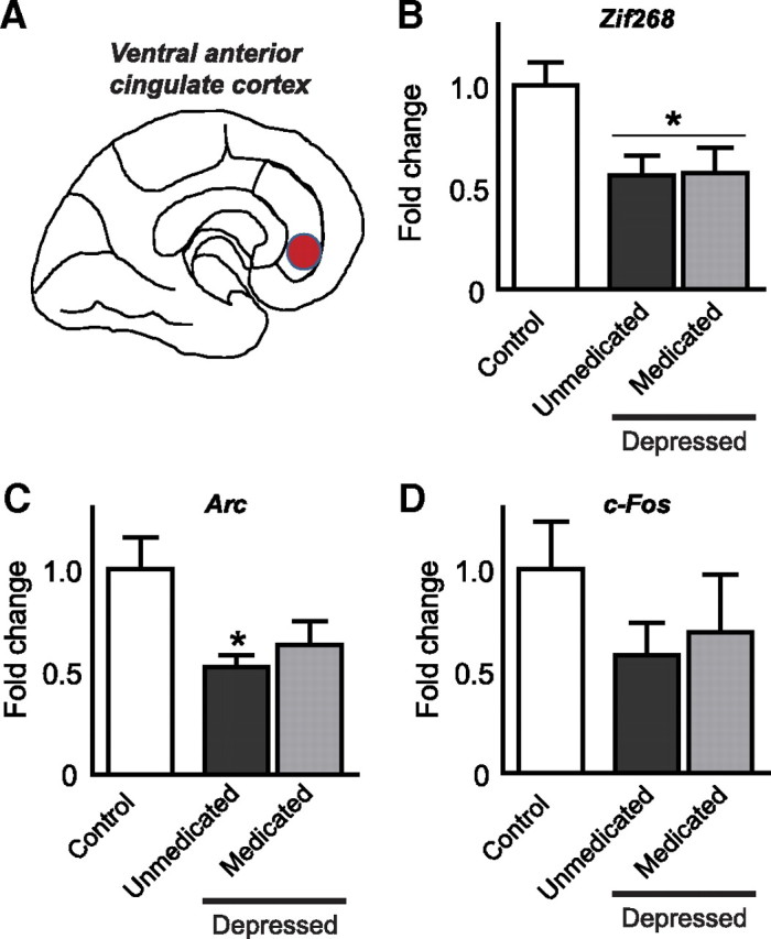
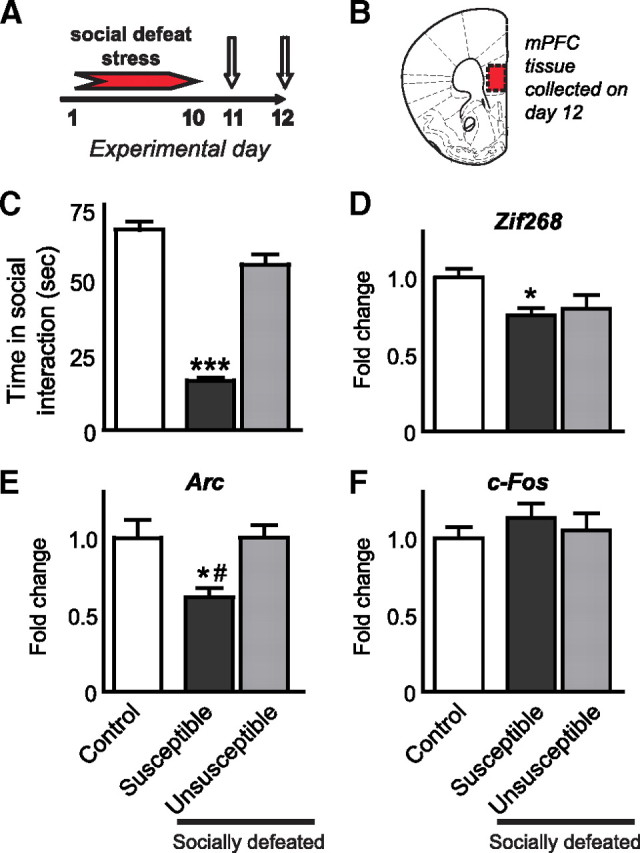
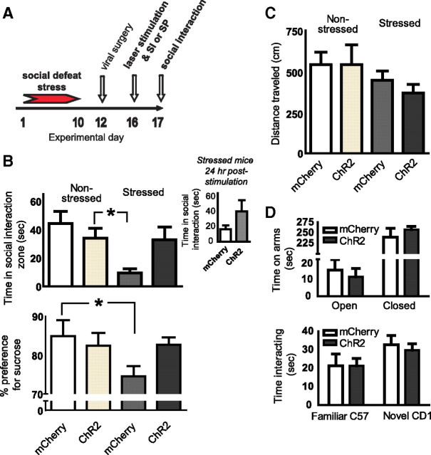
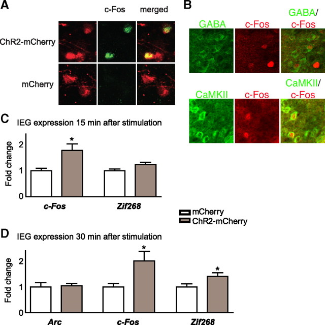
Brain stimulation and imaging studies in humans have highlighted a key role for the prefrontal cortex in clinical depression; however, it remains unknown whether excitation or inhibition of prefrontal cortical neuronal activity is associated with antidepressant responses. Here, we examined cellular indicators of functional activity, including the immediate early genes (IEGs) zif268 (egr1), c-fos, and arc, in the prefrontal cortex of clinically depressed humans obtained postmortem. We also examined these genes in the ventral portion of the medial prefrontal cortex (mPFC) of mice after chronic social defeat stress, a mouse model of depression. In addition, we used viral vectors to overexpress channel rhodopsin 2 (a light-activated cation channel) in mouse mPFC to optogenetically drive "burst" patterns of cortical firing in vivo and examine the behavioral consequences. Prefrontal cortical tissue derived from clinically depressed humans displayed significant reductions in IEG expression, consistent with a deficit in neuronal activity within this brain region. Mice subjected to chronic social defeat stress exhibited similar reductions in levels of IEG expression in mPFC. Interestingly, some of these changes were not observed in defeated mice that escape the deleterious consequences of the stress, i.e., resilient animals. In those mice that expressed a strong depressive-like phenotype, i.e., susceptible animals, optogenetic stimulation of mPFC exerted potent antidepressant-like effects, without affecting general locomotor activity, anxiety-like behaviors, or social memory. These results indicate that the activity of the mPFC is a key determinant of depression-like behavior, as well as antidepressant responses.


Figures

Figure 1.
Figure 1.
A–D, Deficits in IEG expression in the prefrontal cortex of depressed humans. zif268, arc, and c-fos mRNA expression was quantified via qPCR in tissue collected from human postmortem ventral-medial anterior cingulate cortex (A). mRNA levels of zif268 and arc, but not c-fos, were reduced in this region of depressed individuals. These effects predominated in unmedicated depressed individuals, but strong trends were seen in medicated patients who remain symptomatic (B–D). Significant differences from controls are indicated by *p < 0.05.
Figure 2.
Figure 2.
Deficits in IEG expression in mouse mPFC after chronic social defeat stress. zif268, arc and c-fos mRNA expression was quantified via qPCR in tissue collected from mice 48 h after chronic (10 d) social defeat stress. Twenty-four hours after stress, mice were tested for social interaction to identify susceptible and unsusceptible subgroups (A, first arrow). Twenty-four hours after the social interaction test, tissue from the ventral region of the mPFC (B) was collected for IEG analyses in defeated and control mice (A, second arrow). C shows social interaction scores for susceptible and unsusceptible mice. zif268 and arc, but not c-fos, were reduced in the mPFC of stressed mice, and arc was significantly increased in unsusceptible mice compared with susceptible mice (D–F). Significant differences from controls are indicated by *p < 0.05, or ***p < 0.001. Significant differences from unsusceptible are indicated by # p < 0.05.
Figure 3.
Figure 3.
Optogenetic stimulation of mouse mPFC reverses depression-like symptoms induced by chronic social defeat stress. Non-defeated control and socially defeated (susceptible) mice received intra-mPFC injections of viral vectors encoding ChR2-mCherry or mCherry alone. The mPFC was then laser-stimulated to mimic a burst pattern of firing during a test for social interaction (SI) or a test for sucrose preference (SP) (A). During stimulation, stressed mice that express ChR2-mCherry display normative levels of social interaction, unlike defeated mice expressing mCherry only (B, top). Despite a trend, this effect is no longer significant 24 h later when susceptible mice were tested again without additional laser stimulation (B, inset). Social stress-induced deficits in sucrose preference are prevented by mPFC stimulation (B, bottom). General locomotor activity in susceptible and control mice was unaffected by mPFC stimulation in mice expressing either ChR2-mCherry or mCherry alone (C). Optogenetic stimulation of mPFC also does not affect anxiety-like behavior as measured in the elevated plus maze (D), or social memory as observed during a social recognition task (D, bottom). Significant differences between bars are indicated by *p < 0.05.
Figure 4.
Figure 4.
Optogenetic stimulation of the mPFC increases immediate early gene expression. A–D, Laser stimulation induces c-Fos protein specifically in cells that express ChR2 (A). Stimulation-induced c-Fos expression occurs in cells colabeled for CaMKII (i.e., excitatory pyramidal neurons) or GABA (i.e., inhibitory interneurons) (B). Burst patterns of stimulation in the mPFC, after viral expression of ChR2-mCherry, but not mCherry alone, increases cellular measures of functional activity as measured by mRNA expression of c-fos within 15 min of stimulation (C), and both c-fos and zif268, but not arc, within 30 min of stimulation (D). Significant differences between bars are indicated by *p < 0.05.

Similar articles

Cited by

References

    1. Adamantidis AR, Zhang F, Aravanis AM, Deisseroth K, de Lecea L. Neural substrates of awakening probed with optogenetic control of hypocretin neurons. Nature. 2007;450:420–424. - PMC - PubMed
    1. Airan RD, Thompson KR, Fenno LE, Bernstein H, Deisseroth K. Temporally precise in vivo control of intracellular signalling. Nature. 2009;458:1025–1029. - PubMed
    1. Baeg EH, Kim YB, Jang J, Kim HT, Mook-Jung I, Jung MW. Fast spiking and regular spiking neural correlates of fear conditioning in the medial prefrontal cortex of the rat. Cereb Cortex. 2001;11:441–451. - PubMed
    1. Banasr M, Duman RS. Glial loss in the prefrontal cortex is sufficient to induce depressive-like behaviors. Biol Psychiatry. 2008;64:863–870. - PMC - PubMed
    1. Barbas H. Anatomic basis of cognitive-emotional interactions in the primate prefrontal cortex. Neurosci Biobehav Rev. 1995;19:499–510. - PubMed

Publication types