Comparative Study
. 2010 Dec 1;30(48):16091-101.
doi: 10.1523/JNEUROSCI.2400-10.2010.

The Toll-like receptor-3 agonist polyinosinic:polycytidylic acid triggers nigrostriatal dopaminergic degeneration

Affiliations
Comparative Study

The Toll-like receptor-3 agonist polyinosinic:polycytidylic acid triggers nigrostriatal dopaminergic degeneration

Michela Deleidi et al. J Neurosci. .

Abstract

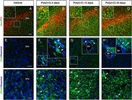
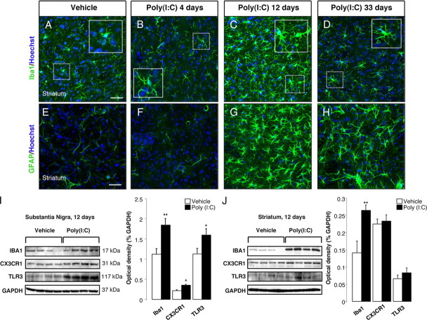
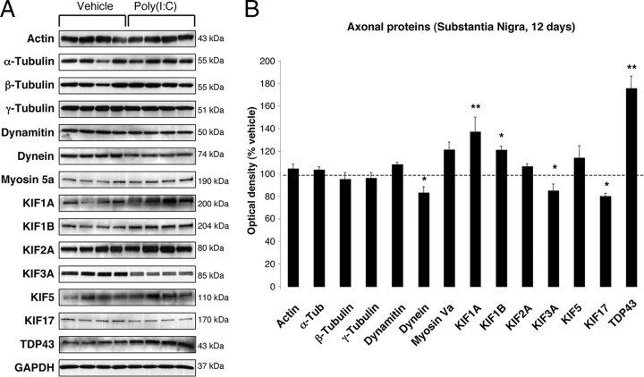
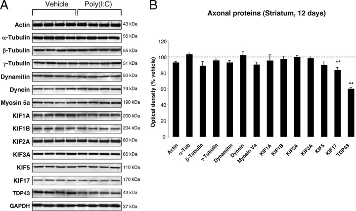
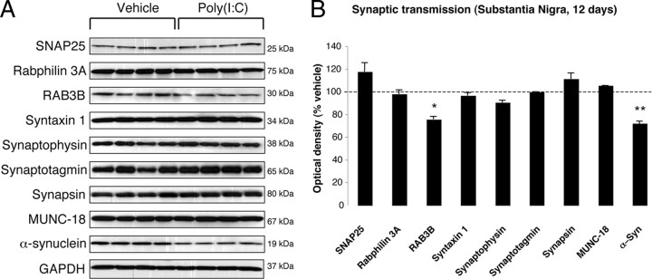
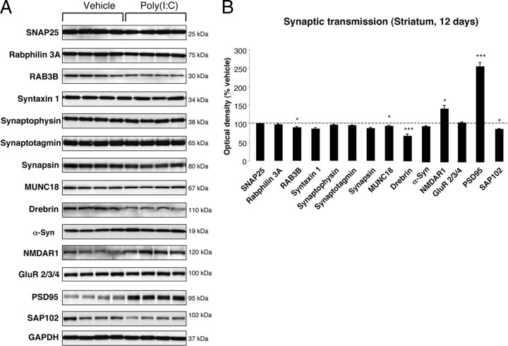
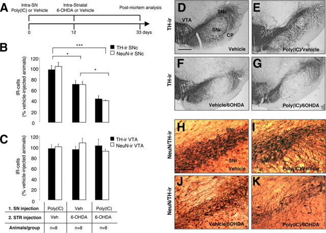
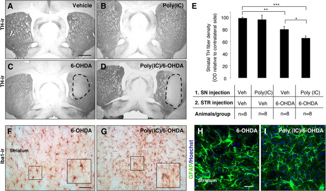
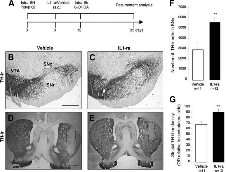
In Parkinson's disease (PD), loss of striatal dopaminergic (DA) terminals and degeneration of DA neurons in the substantia nigra (SN) are associated with glial reactions. Such inflammatory processes are commonly considered an epiphenomenon of neuronal degeneration. However, there is increasing recognition of the role of neuroinflammation as an initiation factor of DA neuron degeneration. To investigate this issue, we established a new model of brain inflammation by injecting the Toll-like receptor 3 (TLR-3) agonist polyinosinic:polycytidylic acid [poly(I:C)] in the SN of adult rats. Poly(I:C) injection induced a sustained inflammatory reaction in the SN and in the dorsolateral striatum. Significant changes were detected in proteins relevant to synaptic transmission and axonal transport. In addition, cytoplasmic mislocalization of neuronal TAR DNA binding protein TDP-43 was observed. Poly(I:C) injection increased the susceptibility of midbrain DA neurons to a subsequent neurotoxic trigger (low-dose 6-hydroxydopamine). Systemic delivery of interleukin-1 receptor antagonist protected SN DA neurons exposed to combined poly(I:C) induced inflammatory and neurotoxic oxidative stress. These data indicate that viral-like neuroinflammation induces predegenerative changes in the DA system, which lowers the set point toward neuronal dysfunction and degeneration. New powerful neuroprotective therapies for PD might be considered by targeting critical inflammatory mechanisms, including cytokine-induced neurotoxicity.


Conflict of interest statement

The authors have no financial conflicts of interest.

Figures

Figure 1.
Figure 1.
Intranigral injection of poly(I:C) induces microglial and astrocytic activation within the SN. Sprague Dawley rats were injected with vehicle or 20 μg of poly(I:C) into the SN. Brains were isolated at 4, 12, and 33 d after injection, and immunofluorescence microscopy was performed for TH (red), Iba1 (green), MHC class II antigen OX6 (green), and GFAP (green). A–D, Iba1 immunohistochemistry revealed resting highly ramified microglia within the SN in vehicle-injected animals (A). In contrast, animals injected with poly(I:C) at 4, 12, and 33 d after the injection showed activated microglia with shorter and thicker processes and larger cell bodies (insets) (B–D). E–H, MHC-II+ cells (arrowheads) were present in poly(I:C)-injected rats at 4,12 and 33 d after injection. I–L, Astrocytic reaction (arrowheads) was found in the SN at 4, 12, and 33 d after poly(I:C) injection. Cell nuclei were counterstained with Hoechst. Dashed lines in G and H indicate blood vessels. Scale bars: A–D, 100 μm; E–L, 50 μm; insets in B–D, 50 μm; insets in F–H, 25 μm. BV, Blood vessel.
Figure 2.
Figure 2.
Intranigral injection of poly(I:C) induces microgliosis and astrocytic activation in the dorsolateral striatum. The dorsolateral striatum was immunostained for Iba1 (green) and GFAP (green) at 4, 12, and 33 d after the intranigral injection of vehicle or 20 μg of poly(I:C). Poly(I:C) promoted microglia activation (A–D) and astrocytic reaction (E–H) within the dorsolateral striatum starting at 12 d after the injection. At day 33, activated astrocytes were still present within the dorsolateral striatum (H), whereas microglial cells showed a resting phenotype (D). Cell nuclei were counterstained with Hoechst. Scale bars: A–H, 50 μm; insets in A–D, 25 μm. I, J, Western blots of SN and striatum extracts from vehicle and poly(I:C)-injected animals. Poly(I:C) injection induced an increase of the levels of Iba1, TLR3, and CX3CR1 in the SN. Levels of Iba1 were increased in the dorsolateral striatum. Vehicle, n = 9; poly(I:C), n = 10; *p < 0.05, **p < 0.01, unpaired t test.
Figure 3.
Figure 3.
Nigral changes in the levels of axonal proteins at 12 d after poly(I:C) injection. A, Anterograde transport motor proteins (KIF1A and KIF1B) were increased, whereas KIF3A, KIF17, and dynein were decreased. Levels of TDP-43 were increased. B, Optical densities of the individual bands were quantified using NIH ImageJ. Optical densities were normalized by the averaged value of GAPDH and expressed as percentage of vehicle-injected conditions. Data are shown as mean ± SEM. Vehicle, n = 9; poly(I:C), n = 10; *p < 0.05, **p < 0.01, unpaired t test.
Figure 4.
Figure 4.
Striatal changes in the levels of axonal proteins at 12 d after poly(I:C) injection. A, Poly(I:C) injection induced decreased levels of KIF17 and TDP-43. B, Optical densities of the individual bands. Data are shown as mean ± SEM. Vehicle, n = 9; poly(I:C), n = 10; **p < 0.01, unpaired t test.
Figure 5.
Figure 5.
Nigral changes in the levels of synaptic proteins at 12 d after poly(I:C) injection. A, Poly(I:C) injection induced decreased levels of RAB3B and α-Syn. B, Optical densities of the individual bands. Data are shown as mean ± SEM. Vehicle, n = 9; poly(I:C), n = 10; *p < 0.05, **p < 0.01, unpaired t test.
Figure 6.
Figure 6.
Striatal changes in the levels of synaptic proteins at 12 d after poly(I:C) injection. A, Poly(I:C) injection induced decreased levels of RAB3B, MUNC18, Drebrin, and SAP102. Levels of the postsynaptic proteins NMDAR1 and PSD-95 were significantly increased. B, Optical densities of the individual bands. Data are shown as mean ± SEM. Vehicle, n = 9; poly(I:C), n = 10; *p < 0.05, ***p < 0.001, unpaired t test.
Figure 7.
Figure 7.
Poly(I:C) injection increases vulnerability to nigral DA neuron loss induced by low-dose 6-OHDA. A, Experimental timeline: rats received a single intranigral injection of 20 μg of poly(I:C) and 12 d later an intrastriatal injection of 5 μg of 6-OHDA. The lesion was allowed to progress for 3 weeks, after which animals were killed for postmortem analyses. B, Unbiased stereological analysis indicates that rats exposed to poly(I:C) injection, followed by a low-dose injection of 6-OHDA, had significant reduction of TH+ neurons in the SNc when compared with animals injected with low-dose 6-OHDA. The loss of TH+ neurons was reflected in the loss of NeuN+ neurons, confirming true neuronal loss. Either vehicle or poly(I:C) injection alone did not induce neuronal cell loss. C, Unbiased stereological analysis indicates no loss of TH+ or NeuN+ neurons in the VTA in any of the groups. D–G, Images of TH immunohistochemistry from single coronal sections of rats injected with vehicle, poly(I:C) alone, 6-OHDA alone, or combined insult. H–K, Images of TH (brown) and NeuN (red) immunohistochemistry from single coronal sections of rats injected with vehicle, poly(I:C) alone, 6-OHDA alone, or combined insult. Scale bar: D–G, 1.0 mm; H–K, 0.25 mm. SNr, Substantia nigra pars reticulata; CP, cerebral peduncle. Animals/group: vehicle/vehicle, n = 8; poly(I:C)/vehicle, n = 8; vehicle/6-OHDA, n = 8; poly(I:C)/6-OHDA, n = 8. *p < 0.05, ***p < 0.001, one-way ANOVA.
Figure 8.
Figure 8.
Intranigral injection of poly(I:C) increases the susceptibility of striatal DA nerve terminals to 6-OHDA toxicity. A–D, Representative striatal sections stained for TH from rats treated with vehicle (A), 20 μg of poly(I:C) (B), 5 μg of 6-OHDA (C), and 20 μg of poly(I:C) followed by 5 μg of 6-OHDA injection (D). E, Optical densitometric quantification of the TH-immunoreactive fibers in the striatum. The injection of 6-OHDA induced a significant loss of TH fibers in the striatum (p ≤ 0.01). The volume of striatal TH+ fibers in the 6-OHDA compared with poly(I:C)/6-OHDA treated rats, expressed relative to the unlesioned side, was significantly higher (p ≤ 0.05). There was no difference between vehicle-treated rats and poly(I:C)-injected rats. F, G, Immunohistochemistry against Iba1 in the striatum of 6-OHDA-lesioned and poly(I:C)/6-OHDA-lesioned rats. Microglia in the striatum of poly(I:C)/6-OHDA-lesioned rats showed activated morphology. H, I, Immunohistochemical staining for GFAP (green) demonstrated similar astrocytic reaction in 6-OHDA-lesioned and poly(I:C)/6-OHDA-lesioned rats. Scale bars: A–D, 2.0 mm; F–I, 50 μm. Animals/group: vehicle/vehicle, n = 8; poly(I:C)/vehicle, n = 8; vehicle/6-OHDA, n = 8; poly(I:C)/6-OHDA, n = 8. *p < 0.05, **p < 0.01, ***p < 0.001, one-way ANOVA.
Figure 9.
Figure 9.
Neutralization of IL-1 reduces cell loss induced by poly(I:C) and 6-OHDA. A, Experimental timeline: rats received a single intranigral injection of 20 μg of poly(I:C) and 9 d later started on either IL-1ra or vehicle, which continued through the experiment. All animals received an intrastriatal injection of 5.0 μg of 6-OHDA 12 d after vehicle/poly(I:C) injection and were allowed 21 d until postmortem analyses. B–E, Representative nigral and striatal sections stained for TH from rats treated with vehicle (B, D) or IL-1ra (C, E). F, Stereological quantification TH+ cells in the SNc revealed that IL-1ra prevented dopaminergic cell loss associated with poly(I:C) and 6-OHDA. G, Optical densitometric quantification of the TH-immunoreactive fibers in the striatum revealed that IL-1ra prevented TH fiber loss in the striatum. Data are shown as mean ± SEM. Vehicle, n = 11; IL-1ra, n = 12. **p < 0.01, unpaired t test. Scale bars: B, C, 0.25 mm; D, E, 1.0 mm. SNr, Substantia nigra pars reticulata.

References

    1. Alexopoulou L, Holt AC, Medzhitov R, Flavell RA. Recognition of double-stranded RNA and activation of NF-kappaB by Toll-like receptor 3. Nature. 2001;413:732–738. - PubMed
    1. Allan SM, Tyrrell PJ, Rothwell NJ. Interleukin-1 and neuronal injury. Nat Rev Immunol. 2005;5:629–640. - PubMed
    1. Aoki C, Sekino Y, Hanamura K, Fujisawa S, Mahadomrongkul V, Ren Y, Shirao T. Drebrin A is a postsynaptic protein that localizes in vivo to the submembranous surface of dendritic sites forming excitatory synapses. J Comp Neurol. 2005;483:383–402. - PubMed
    1. Banks GT, Fisher EM. Cytoplasmic dynein could be key to understanding neurodegeneration. Genome Biol. 2008;9:214. - PMC - PubMed
    1. Berth SH, Leopold PL, Morfini GN. Virus-induced neuronal dysfunction and degeneration. Front Biosci. 2009;14:5239–5259. - PubMed

Publication types

LinkOut - more resources