Comparative Study
. 2010 Dec 1;30(48):16188-96.
doi: 10.1523/JNEUROSCI.2265-10.2010.

The prefrontal cortex communicates with the amygdala to impair learning after acute stress in females but not in males

Affiliations
Comparative Study

The prefrontal cortex communicates with the amygdala to impair learning after acute stress in females but not in males

Lisa Y Maeng et al. J Neurosci. .

Abstract

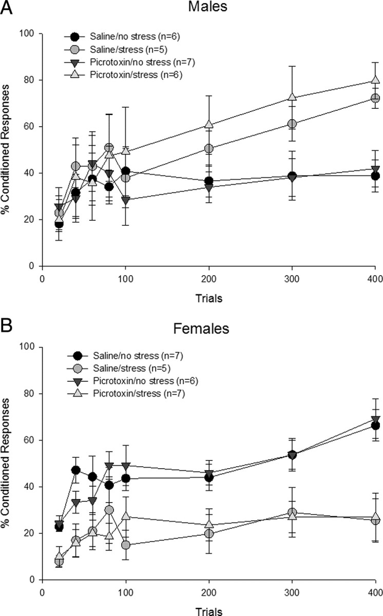
Acute stress exposure enhances classical eyeblink conditioning in male rats, whereas exposure to the same event dramatically impairs performance in females (Wood and Shors, 1998; Wood et al., 2001). We hypothesized that stress affects learning differently in males and females because different brain regions and circuits are being activated. In the first experiment, we determined that neuronal activity within the medial prefrontal cortex (mPFC) during the stressful event is necessary to disrupt learning in females. In both males and females, the mPFC was bilaterally inactivated with GABA agonist muscimol before the stressor. Inactivation prevented only the impaired performance in females; it had no consequence for performance in males. However, in the second experiment, excitation of the mPFC alone with GABA antagonist picrotoxin was insufficient to elicit the stress effect that was prevented through the inactivation of this region in females. Therefore, we hypothesized that the mPFC communicates with the basolateral amygdala to disrupt learning in females after the stressor. To test this hypothesis, these structures were disconnected from each other with unilateral excitotoxic (NMDA) lesions on either the same or opposite sides of the brain. Females with contralateral lesions, which disrupt the connections on both sides of the brain, were able to learn after the stressful event, whereas those with ipsilateral lesions, which disrupt only one connection, did not learn after the stressor. Together, these data indicate that the mPFC is critically involved in females during stress to impair subsequent learning and does so via communication with the amygdala.


Figures

Figure 1.
Figure 1.
Histology of mPFC cannulation. Cannula tip placements within the mPFC were between +3.20 and +2.70 mm relative to bregma. Animals were included if tips of the injection cannula were within the dorsal boundary of the prelimbic cortex and at least 1 mm above the ventral boundary of the infralimbic cortex. Photograph of mPFC section stained with 0.1% neutral red with cannula tip placements marked by Prussian blue is also included. p, Prelimbic region of the medial prefrontal cortex; I, infralimbic region of the medial prefrontal cortex.
Figure 2.
Figure 2.
mPFC-BLA disconnection procedure. Animals with contralateral excitotoxic lesions received a unilateral lesion to the mPFC and a unilateral lesion to the BLA in opposite hemispheres. The contralateral lesion disrupted communication between the mPFC and BLA in both hemispheres. Animals with ipsilateral lesions received a unilateral lesion of the mPFC and a unilateral lesion of the BLA within the same hemisphere. Thus, the connection between the two structures was preserved in one hemisphere in these rats. If the concurrent activation of both the mPFC and BLA is necessary for the learning deficit after stress, then animals with contralateral lesions should learn well even though they were exposed to the stressor.
Figure 3.
Figure 3.
Histology of mPFC-BLA disconnection. A, mPFC lesions. B, BLA lesions. Largest lesions (in gray) and smallest lesions (in black) of rats included are depicted here. The unilateral images are representative of lesions in both hemispheres. Brain sections of the mPFC and BLA were stained with 0.1% cresyl violet to verify sites of excitotoxic damage (marked by arrowheads). Note the darkly stained astrocytes and absence of cell bodies in lesioned tissue (4×). C, Intact mPFC. D, Lesioned mPFC. E, Lesioned BLA. F, Intact BLA.
Figure 4.
Figure 4.
mPFC inactivation. The mPFC was inactivated with muscimol or infused with aCSF during the stressor. One day later, animals were trained with delay conditioning. A, Males treated with muscimol and stressed emitted more CRs than those treated with either vehicle or muscimol and not stressed. They performed similarly to those that were stressed and infused with aCSF. Thus, inactivating the mPFC did not prevent the stress-induced facilitation of learning in males. B, Stressed females infused with vehicle expressed fewer CRs than their unstressed counterparts. However, when females were infused with muscimol during the stressor, they increased responding and performed similarly to those that were unstressed. Thus, in contrast to males, mPFC inactivation in females eliminated the modified behavioral response after stress.
Figure 5.
Figure 5.
mPFC activation. The mPFC was infused with picrotoxin or saline during the stressor. One day later, animals were trained with delay conditioning. A, Picrotoxin-treated stressed males emitted more CRs than the saline-treated unstressed males but performed similarly to those that were stressed and infused with saline. Thus, activating the mPFC did not alter the stress-induced facilitation of learning or performance in the absence of stress in males. B, Stressed females infused with saline expressed fewer CRs than their unstressed counterparts. Picrotoxin-infused stressed females performed similarly to the saline-treated stressed females, emitting fewer CRs than picrotoxin-treated unstressed animals. Thus, mPFC activation in females had no effect on decremented conditioned responding following stress exposure or performance in the absence of stress.
Figure 6.
Figure 6.
mPFC-BLA disconnection. Acute stressful experience disrupted learning in females with ipsilateral mPFC-BLA lesions. In contrast, conditioned responding of animals with contralateral lesions was not impaired by stress and was similar to the performance of the unstressed females with both types of lesions. Thus, communication between the mPFC and BLA is necessary to impair learning after a stressful event in females.
Figure 7.
Figure 7.
The percentage of female rats in each lesion and stress condition that met learning criterion. Animals that learned emitted at least 60% CRs in at least two (of the four) consecutive sessions of 100 trials.

References

    1. Amat J, Baratta MV, Paul E, Bland ST, Watkins LR, Maier SF. Medial prefrontal cortex determines how stressor controllability affects behavior and dorsal raphe nucleus. Nat Neurosci. 2005;8:365–371. - PubMed
    1. Bacon SJ, Headlam AJ, Gabbott PL, Smith AD. Amygdala input to medial prefrontal cortex (mPFC) in the rat: a light and electron microscope study. Brain Res. 1996;720:211–219. - PubMed
    1. Bangasser DA, Shors TJ. Acute stress impairs trace eyeblink conditioning in females without altering the unconditioned response. Neurobiol Learn Mem. 2004;82:57–60. - PMC - PubMed
    1. Bangasser DA, Shors TJ. The hippocampus is necessary for enhancements and impairments of learning following stress. Nat Neurosci. 2007;10:1401–1403. - PMC - PubMed
    1. Bangasser DA, Shors TJ. The bed nucleus of the stria terminalis modulates learning after stress in masculinized but not cycling females. J Neurosci. 2008;28:6383–6387. - PMC - PubMed

Publication types