

doi: 10.2106/JBJS.J.00780.
Effects of construct stiffness on healing of fractures stabilized with locking plates
Affiliations
- PMID: 21123589
- PMCID: PMC2995582
- DOI: 10.2106/JBJS.J.00780
Item in Clipboard
Effects of construct stiffness on healing of fractures stabilized with locking plates
J Bone Joint Surg Am.
2010 Dec.
No abstract available
Figures
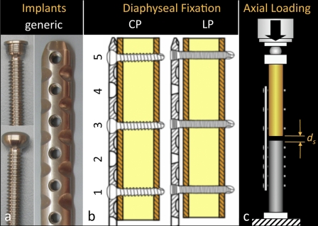
The stiffness and interfragmentary motion of locked-plate constructs was assessed. a: Generic 4.5-mm plate and screws. b: Diaphyseal fixation with a conventional plate (CP) or a locking plate (LP). c: Axial loading and assessment of resulting interfragmentary motion (ds) at the near and far cortices.
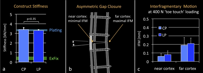
a: Conventional (CP) and locked-plate (LP) constructs were comparably stiff and were approximately one order of magnitude stiffer than external fixators (ExFix)-. b: Axial loading caused plate bending and asymmetric gap closure, whereby interfragmentary motion (IFM) at the near cortex was minimal. c: Near-cortex motion in response to 400-N simulated toe-touch loading remained below 0.1 mm, which is considered insufficient to promote secondary bone-healing.
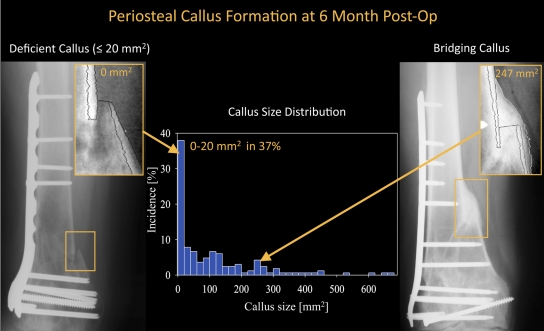
Periosteal callus measurement at six months postoperatively in a study of patients yielded examples of deficient callus formation (≤20 mm2) and bridging callus (247 mm2). The callus size distribution illustrates that 37% of all fractures had formed no or very little callus (≤20 mm2) at six months.
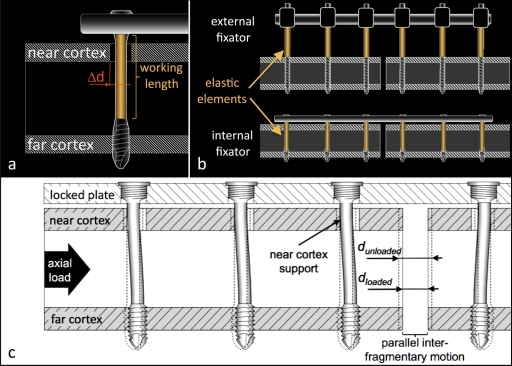
Far-cortical-locking concept. a: Far-cortical-locking screws lock into the plate and the far cortex. The screws have a reduced midshaft diameter to retain a controlled motion envelope (Δd) in the near cortex, which increases the screw's working length. b: Analogous to external fixators, far-cortical-locking constructs derive a low stiffness from elastic flexion of the screw shafts. c: Flexion of far-cortical-locking screws within the near-cortex motion envelope induces parallel motion at the fracture gap.
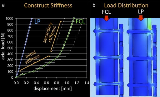
a: Far-cortical-locking (FCL) constructs exhibit a biphasic stiffness profile, similar to that of Ilizarov external fixators. The primary stiffness was 88% lower than that of the standard locked-plate (LP) construct, enabling interfragmentary motion at reduced postoperative loading. At elevated loads, far-cortical-locking stiffness increases as a result of the additional support of screws at the near cortex. (Reprinted from: Bottlang M, Doornink J, Fitzpatrick DC, Madey SM. Far cortical locking can reduce stiffness of locked-plate constructs while retaining construct strength. J Bone Joint Surg Am. 2009;91:1988.) b: The elastic shaft of far-cortical-locking screws allows evenly distributed load sharing between screws and effectively prevents the stress risers seen at the end-screw of standard locked-plate constructs.
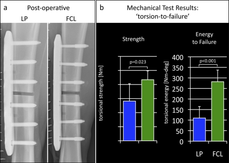
Evaluation of fracture-healing with locked-plate (LP) and far-cortical-locking (FCL) constructs in an ovine fracture-healing model. a: Postoperative radiographs depicting 3-mm-gap tibial osteotomy sites stabilized with locked-plate and far-cortical-locking constructs. b: Tibiae treated with far-cortical-locking constructs healed to be 54% stronger with torsional testing and tolerated 156% more energy to failure than tibiae treated with standard locking plates.
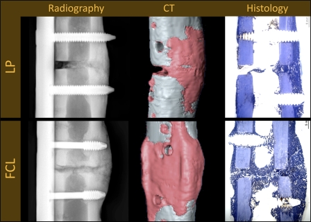
Radiographic, computed tomography (CT), and histological evaluation of the tibiae after the sheep was killed at nine weeks after surgery. The locked-plate (LP) constructs suppressed callus formation at the near cortex, where gap motion is minimal, leading to partial nonunions in three of the six sheep. The far-cortical-locking (FCL) constructs induced more callus formation, symmetric callus, and reliable bridging in all six sheep.
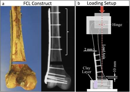
a: Periarticular far-cortical-locking (FCL) construct, whereby far-cortical-locking screws are used only in the diaphyseal segment and metaphyseal fixation is performed with a standard locked-plate technique. b: Quasi-physiologic loading of cadaveric femora along the mechanical axis to assess construct stiffness, durability, and residual strength was done with this setup.

a: In response to loading at one times body weight, standard periarticular locking constructs (the locked-plate [LP] group) induced asymmetric fracture motion. Near-cortex motion remained below 0.1 mm, which is considered insufficient to promote secondary bone-healing. Under the same load, far-cortical-locking (FCL) constructs induced essentially parallel motion, sufficient to promote secondary bone-healing. b: While far-cortical-locking screws reduced construct stiffness by 81%, the far-cortical-locking constructs remained as strong as the locked-plate constructs.
References
-
- Kubiak EN, Fulkerson E, Strauss E, Egol KA. The evolution of locked plates. J Bone Joint Surg Am. 2006;88 Suppl 4:189-200 - PubMed
-
- Ring D, Kloen P, Kadzielski J, Helfet D, Jupiter JB. Locking compression plates for osteoporotic nonunions of the diaphyseal humerus. Clin Orthop Relat Res. 2004;425:50-4 - PubMed
-
- Perren SM. Evolution of the internal fixation of long bone fractures. The scientific basis of biological internal fixation: choosing a new balance between stability and biology. J Bone Joint Surg Br. 2002;84:1093-110 - PubMed
-
- Tan SL, Balogh ZJ. Indications and limitations of locked plating. Injury. 2009;40:683-91 - PubMed
Publication types
MeSH terms
Grants and funding
LinkOut - more resources
Full Text Sources
Other Literature Sources
Medical
