Review
. 2011 Jan;4(1):31-41.
doi: 10.1242/dmm.006403. Epub 2010 Dec 1.

Degeneration and regeneration of the intervertebral disc: lessons from development

Affiliations
Review

Degeneration and regeneration of the intervertebral disc: lessons from development

Lachlan J Smith et al. Dis Model Mech. 2011 Jan.

Abstract

Degeneration of the intervertebral discs, a process characterized by a cascade of cellular, biochemical, structural and functional changes, is strongly implicated as a cause of low back pain. Current treatment strategies for disc degeneration typically address the symptoms of low back pain without treating the underlying cause or restoring mechanical function. A more in-depth understanding of disc degeneration, as well as opportunities for therapeutic intervention, can be obtained by considering aspects of intervertebral disc development. Development of the intervertebral disc involves the coalescence of several different cell types through highly orchestrated and complex molecular interactions. The resulting structures must function synergistically in an environment that is subjected to continuous mechanical perturbation throughout the life of an individual. Early postnatal changes, including altered cellularity, vascular regression and altered extracellular matrix composition, might set the disc on a slow course towards symptomatic degeneration. In this Perspective, we review the pathogenesis and treatment of intervertebral disc degeneration in the context of disc development. Within this scope, we examine how model systems have advanced our understanding of embryonic morphogenesis and associated molecular signaling pathways, in addition to the postnatal changes to the cellular, nutritional and mechanical microenvironment. We also discuss the current status of biological therapeutic strategies that promote disc regeneration and repair, and how lessons from development might provide clues for their refinement.


Figures

Fig. 1.
Fig. 1.
Schematic representations of the adult intervertebral disc. (A) Mid-sagittal cross-section showing anatomical regions. (B) Three-dimensional view illustrating AF lamellar structure.
Fig. 2.
Fig. 2.
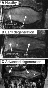
Magnetic resonance images illustrating different stages of human lumbar disc degeneration. (A) A healthy disc exhibiting distinct AF lamellae (AF) and central NP region (NP). (B) A disc exhibiting early stages of degeneration, including moderate height reduction, decreased NP signal intensity and inward bulging of AF lamellae (*). (C) A disc exhibiting advanced stages of degeneration, including severely reduced height, large fissure (*) and generalized structural deterioration. Images obtained using 7T Siemens scanner and a turbo spin echo sequence at 200 μm isotropic voxel resolution.
Fig. 3.
Fig. 3.
Schematic representation of embryonic morphogenesis of the mammalian intervertebral disc. Colors represent origins and fates of cell populations. Also indicated are key morphogens and transcriptional regulators implicated in the growth and differentiation of the disc structures at each developmental stage. (A) The notochord adjacent to pairs of paraxial somites, which contain sclerotome cells. (B) Sclerotome cells condense around the notochord. (C) Cells adopt a metameric pattern of more condensed (green) and less condensed (brown) regions that give rise to the disc and vertebral bodies, respectively. (D) The notochord contracts within the vertebral body rudiments and expands within the future intervertebral disc to form the NP. (E) Basic structures of the disc are established, and AF cells adopt orientations and alignments that form the template for the lamellar structure. VB, vertebral body.
Fig. 4.
Fig. 4.
Stages of notochord transformation into the NP in the mouse embryo. Embryos were stained with Alcian Blue (which marks glycosaminoglycans) and Picrosirius Red (which marks collagen). At embryonic day (E)12.5, the notochord (arrow) runs uninterrupted the length of the vertebral column. Sclerotome cells have condensed perichordially, and metameric patterning of future disc and vertebral body condensations is apparent. At E13.5, the notochord has begun to contract within the vertebral body regions and expand within the disc regions (arrow). At E14.5, the notochord has virtually disappeared from the vertebral bodies, persisting solely in the locations of the future NPs (arrow). In the neonate, lateral expansion of the NP has occurred (arrow) and primary ossification centers are present in the vertebral bodies. At postnatal age 1 month, vertebral bodies are fully ossified and the NP (arrow) contains a glycosaminoglycan-rich extracellular matrix surrounded by the collagenous AF. Scale bars: E12.5, E13.5 and E14.5: 200 μm (top) and 100 μm (bottom); neonate: 400 μm (top) and 200 μm (bottom); 1 month: 500 μm (top) and 400 μm (bottom).

References

    1. Acaroglu E.R., Iatridis J.C., Setton L.A., Foster R.J., Mow V.C., Weidenbaum M. (1995). Degeneration and aging affect the tensile behavior of human lumbar anulus fibrosus. Spine 20, 2690–2701 - PubMed
    1. Adams D.S., Keller R., Koehl M.A. (1990). The mechanics of notochord elongation, straightening and stiffening in the embryo of Xenopus laevis. Development 110, 115–130 - PubMed
    1. Adams M.A., Roughley P.J. (2006). What is intervertebral disc degeneration, and what causes it? Spine 31, 2151–2161 - PubMed
    1. Adams M.A., McNally D.S., Dolan P. (1996). ‘Stress’ distributions inside intervertebral discs. The effects of age and degeneration. J. Bone Joint Surg. Br. 78, 965–972 - PubMed
    1. Aguiar D.J., Johnson S.L., Oegema T.R. (1999). Notochordal cells interact with nucleus pulposus cells: regulation of proteoglycan synthesis. Exp. Cell Res. 246, 129–137 - PubMed

Publication types

MeSH terms