. 2010 Dec 1;24(23):2666-77.
doi: 10.1101/gad.1964410.

Inhibition of RIP2's tyrosine kinase activity limits NOD2-driven cytokine responses

Affiliations

Inhibition of RIP2's tyrosine kinase activity limits NOD2-driven cytokine responses

Justine T Tigno-Aranjuez et al. Genes Dev. .

Abstract

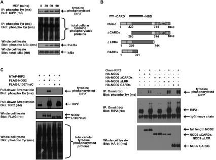
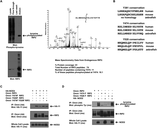
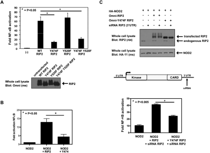
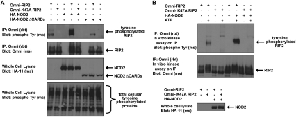
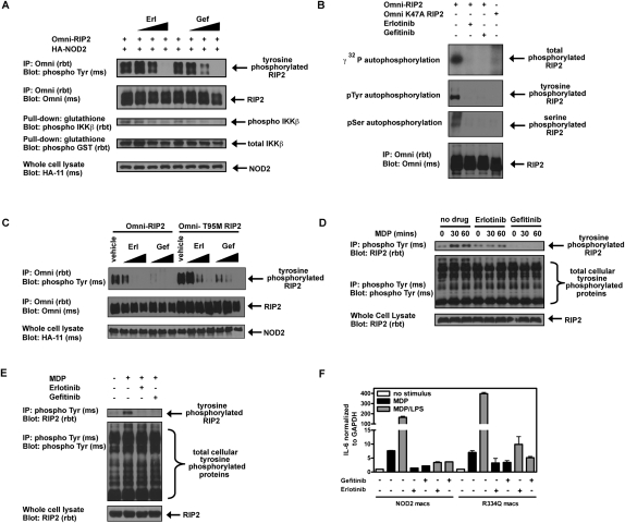
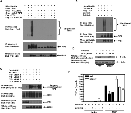
Upon intracellular bacterial exposure, the Crohn's disease and sarcoidosis susceptibility protein NOD2 (nucleotide oligomerization domain protein 2) binds to the protein kinase RIP2 (receptor-interacting protein 2) to coordinate NF-κB (nuclear factor κ B)-mediated cytokine responses. While RIP2 clearly has kinase activity, the function of its kinase domain has been enigmatic. Although originally classified as a serine-threonine kinase based on homology scans, we find that RIP2 also has tyrosine kinase activity. RIP2 undergoes autophosphorylation on Tyr 474 (Y474). This phosphorylation event is necessary for effective NOD2 signaling and does not occur in the presence of the most common Crohn's disease-associated NOD2 allele. Given this tyrosine kinase activity, a small-molecule inhibitor screen designed to identify pharmacologic agents that inhibit RIP2's tyrosine kinase activity was performed. At nanomolar concentrations, the EGFR (epidermal growth factor receptor) tyrosine kinase inhibitors gefitinib (Iressa) and erlotinib (Tarceva) were found to inhibit both RIP2 tyrosine phosphorylation and MDP (muramyl dipeptide)-induced cytokine release in a variety of NOD2 hyperactivation states. This effect is specific for RIP2 and does not depend on EGFR. The finding that RIP2 has tyrosine kinase activity and the finding that gefitinib and erlotinib, two agents already used clinically for cancer chemotherapy, can inhibit this activity suggest that RIP2's tyrosine kinase activity could be targeted specifically in the treatment of inflammatory diseases.


Figures

Figure 1.
Figure 1.
RIP2 is tyrosine-phosphorylated in response to NOD2 activation. (A) HT-29 cells were stimulated with 10 μg/mL MDP, and lysates were harvested at the indicated time points. Tyrosine-phosphorylated proteins were immunoprecipitated, and Western blotting was performed using the indicated antibodies. NOD2 activation stimulated RIP2 tyrosine phosphorylation. (B) HEK293 cells were transfected with wild-type NOD2 or with NOD2 deletion mutants as shown in the top schematic. RIP2 was immunoprecipitated, and Western blotting was performed using the indicated antibodies. NOD2-induced RIP2 tyrosine phosphorylation required the presence of the CARDs of NOD2, indicating that NOD2:RIP2 binding was essential. (C) HEK293 cells were transfected with the indicated constructs. RIP2 was immunoprecipitated and Western blotting was performed. RIP2 tyrosine phosphorylation is lost with the loss-of-function Crohn's disease-associated NOD2 polymorphism L1007insC.
Figure 2.
Figure 2.
RIP2 is phosphorylated on Tyr 474. (A) HT-29 cells were treated with 10 μg/mL MDP for 45 min. Lysates were generated and endogenous RIP2 was immunoprecipitated. Western blotting showed that RIP2 was purified from the lysate (bottom panel), and that this contained extensive tyrosine phosphorylation (top panel). This tyrosine-phosphorylated RIP2 was excised from the gel and subjected to MS analysis. Overall, in this experiment, 74 peptides corresponding to RIP2 were identified and 61% protein coverage was obtained. The MS/MS spectrum of the RIP2 peptide DLIMKEDpYELVSTKPTR showing a single phosphorylation site at Tyr 8. Fragment ions are consistent with the phosphorylation site localized to Tyr 8 in the above peptide. The phosphate loss at b8 proves that phosphorylation is at Tyr 8, as does the lack of phosphate losses from y1 through y9. However, phosphate losses at y12 and y15 also add credibility to phosphorylation at Tyr 8. These findings show that phosphorylation can occur at Y474 in endogenous RIP2. (B) Additional MS analysis by our laboratory and others (Daub et al. 2008; Oppermann et al. 2009) (in total covering collectively 90% of the RIP2 protein) found a total of three tyrosine phosphorylation sites on RIP2 (Y381, Y474, and Y527). Conservation of these sites is shown. Y474 and Y527 are conserved in zebrafish, while Y381 is not conserved. (C) HA-tagged NOD2 was coexpressed with RIP2, Y474F RIP2, Y520F RIP2, or Y474FY520F RIP2. Immunoprecipitations showed that all four RIP2 proteins could coprecipitate with NOD2. (D) HEK293 cells were transfected with or without NOD2 in the presence of wild-type (WT), Y474F, or Y520F RIP2. RIP2 was immunoprecipitated, and Western blotting was performed using the indicated antibodies. Mutation of Tyr 474 but not Tyr 520 on RIP2 inhibits NOD2-induced RIP2 tyrosine phosphorylation.
Figure 3.
Figure 3.
Mutation of Y474 causes a decrease in RIP2's ability to induce NF-κB. (A) NF-κB luciferase assays were performed to measure RIP2 or the RIP2 mutants' ability to activate NF-κB. Mutation of Tyr 474 but not Tyr 520 strongly inhibited RIP2's ability to activate NF-κB (P < 0.03, N = 4). (B) A similar experiment was performed in RIP2−/− MEFs. NF-κB luciferase assays showed that, in these cells, NOD2 could not activate NF-κB without the presence of wild-type RIP2. Activation with NOD2 and Y474F RIP2 was significantly lower (P < 0.015, N = 5). (C) To determine the effect of loss of wild-type, endogenous RIP2 expression on NOD2-induced NF-κB activity, endogenous RIP2 expression was inhibited through the use of an siRNA that targets the 3′UTR of RIP2. (Top panels) Western blotting showed that, while endogenous RIP2 could be silenced using this siRNA, the cDNA constructs expressing either wild-type RIP2 or Y474F RIP2 (both lacking the 3′UTR) could rescue the expression. NF-κB-driven luciferase assays were then performed to determine the synergy between NOD2 and either RIP2 or Y474F RIP2. NOD2 cDNA transfection expression was titrated (75 ng) to a dose in which we found synergy with RIP2 (350 ng) to be maximal, and NF-κB luciferase assays were performed in the presence of the siRNA targeting endogenous RIP2. Synergy with Y474F RIP2 was significantly lower when compared with wild-type RIP2 (P < 0.005, N = 4).
Figure 4.
Figure 4.
RIP2 autophosphorylates on tyrosine. (A) HEK293 cells were transfected with NOD2 in the presence of wild-type or K47A (kinase-inactive) RIP2. RIP2 was immunoprecipitated and Western blotting was performed. RIP2 kinase activity is required for NOD2-induced tyrosine phosphorylation of RIP2. (B) HEK293 cells were transfected with the indicated constructs, and RIP2 was immunoprecipitated under stringent washing conditions. Purified RIP2 or kinase-dead RIP2 (K47A) was then incubated with or without ATP in an in vitro kinase assay. Addition of SDS-PAGE sample buffer followed by boiling stopped the reaction. Western blotting was then performed using the indicated antibodies. RIP2 kinase activity is required for in vitro tyrosine autophosphorylation.
Figure 5.
Figure 5.
Erlotinib (Tarceva) and gefitinib (Iressa) inhibit RIP2 tyrosine phosphorylation. (A) HEK293 cells were transfected with RIP2, IKKβ, and NOD2. Lysates were generated and immunoprecipitations were performed. Western blotting was then performed using the indicated antibodies. Increasing doses of erlotinib and gefitinib (10 nM, 100 nM, and 1 μM) inhibited RIP2 tyrosine phosphorylation, and this inhibition matched the inhibition of RIP2-induced IKKβ activation. (B) To determine whether gefitinib and erlotinib could inhibit RIP2's kinase activity in vitro and whether these compounds inhibited both serine–threonine and tyrosine kinase activity of RIP2, in vitro kinase assays were performed using either immunoprecipitated RIP2 or kinase-dead (K47A) RIP2. One-hundred nanomolar gefitinib and 100 nM erlotinib could inhibit total autophosphorylation activity of RIP2 (32P incorporation, top autoradiograph) and autophosphorylation on both tyrosines and serines, as shown by Western blotting with anti-phosphoserine and anti-phosphotyrosine antibodies. (C) HEK293 cells were transfected with NOD2 and wild-type or T95M RIP2 for 6 h before treatment with erlotinib and gefitinib (100 nM, 500 nM, and 2 μM). The T95M mutation is homologous to the EGFR erlotinib and gefitinib-resistant mutation (T790M) that occurs in resistant cancer patients. After 16 h, lysates were harvested for immunoprecipitation and Western blotting. Erlotinib and gefitinib inhibited RIP2 tyrosine phosphorylation in a dose-dependent manner and are potent at nanomolar concentrations. The sensitivity decreased at least fivefold when T95M RIP2 was used. (D,E) HT-29 cells overexpressing NOD2 (D) or RAW264.7 macrophages overexpressing NOD2 (E) either were not pretreated or were pretreated with erlotinib and gefitinib (500 nM). One hour later, the cells were stimulated with 10 μg/mL MDP for the time points indicated before harvesting lysates for immunoprecipitation and Western blotting. While there was basal tyrosine phosphorylation of RIP2 in untreated cells, both erlotinib and gefitinib inhibit MDP-induced RIP2 tyrosine phosphorylation in both these divergent cell lines. (F) RAW264.7 macrophages overexpressing wild-type NOD2 or the activating R334Q NOD2 mutation associated with Blau Syndrome were pretreated with erlotinib and gefitinib for 1 h before stimulation with MDP (10 μg/mL) or MDP (10 μg/mL) + LPS (5 ng/mL) . Sixteen hours later, cells were harvested to obtain RNA for RT–PCR analysis. Erlotinib and gefitinib inhibit the NOD2–TLR synergy seen in either wild-type NOD2 cells or R334Q Blau Syndrome NOD2 cells. This finding was also true for TNF-α (not shown).
Figure 6.
Figure 6.
Tyrosine phosphorylation of RIP2 promotes its ubiquitination by the E3 ligase ITCH, and inhibition of this tyrosine phosphorylation inhibits the exaggerated MDP responses seen in ITCH−/− cells. (A) HEK293 cells were transfected with the indicated constructs. RIP2 was immunoprecipitated, and immunoblotting with the indicated antibodies was performed. Both the kinase activity of RIP2 and phosphorylation at Tyr 474 are required for ITCH-induced ubiquitination of RIP2. (B) HEK293 cells were transfected with the indicated constructs. Six hours later, cells were treated with erlotinib and gefitinib overnight before harvesting cell lysates for immunoprecipitation. Western blotting showed that both erlotinib and gefitinib inhibit ITCH-induced ubiquitination of RIP2. (C) HEK293 cells were transfected with RIP2 and NOD2 in the absence or presence of four different siRNAs against ITCH. Cell lysates were harvested 24 h after transfection for immunoprecipitation and Western blotting. siRNA knockdown of ITCH enhances NOD2-induced RIP2 tyrosine phosphorylation, suggesting that ITCH ubiquitinates and down-regulates the tyrosine-phosphorylated form of RIP2. (D) BMDMs from wild-type or ITCH−/− mice were treated with gefitinib overnight before stimulation with 10 μg/mL MDP. Cell lysates were collected at the time points indicated for immunoprecipitation and Western blotting. Gefitinib inhibits MDP-induced NF-κB activation in both wild-type and ITCH−/− mice. (E) BMDMs from wild-type or ITCH−/− mice were treated with erlotinib and gefitinib for 1 h before stimulation with 10 μg/mL MDP. After 24 h, supernatants were harvested for TNF-α ELISA. Erlotinib and gefitinib can reduce the exacerbated MDP-induced cytokine responses in the ITCH−/− mice to wild-type levels.

References

    1. Abbott DW, Wilkins A, Asara JM, Cantley LC 2004. The Crohn's disease protein, NOD2, requires RIP2 in order to induce ubiquitinylation of a novel site on NEMO. Curr Biol 14: 2217–2227 - PubMed
    1. Abbott DW, Yang Y, Hutti JE, Madhavarapu S, Kelliher MA, Cantley LC 2007. Coordinated regulation of Toll-like receptor and NOD2 signaling by K63-linked polyubiquitin chains. Mol Cell Biol 27: 6012–6025 - PMC - PubMed
    1. Argast GM, Fausto N, Campbell JS 2005. Inhibition of RIP2/RIck/CARDIAK activity by pyridinyl imidazole inhibitors of p38 MAPK. Mol Cell Biochem 268: 129–140 - PubMed
    1. Barnich N, Aguirre JE, Reinecker HC, Xavier R, Podolsky DK 2005. Membrane recruitment of NOD2 in intestinal epithelial cells is essential for NF-κB activation in muramyl dipeptide recognition. J Cell Biol 170: 21–26 - PMC - PubMed
    1. Bertrand MJ, Doiron K, Labbe K, Korneluk RG, Barker PA, Saleh M 2009. Cellular inhibitors of apoptosis cIAP1 and cIAP2 are required for innate immunity signaling by the pattern recognition receptors NOD1 and NOD2. Immunity 30: 789–801 - PubMed

Publication types

MeSH terms