. 2011 Mar;105(3):1051-62.
doi: 10.1152/jn.00831.2010. Epub 2010 Dec 1.

CB1 modulation of temporally distinct synaptic facilitation among local circuit interneurons mediated by N-type calcium channels in CA1

Affiliations

CB1 modulation of temporally distinct synaptic facilitation among local circuit interneurons mediated by N-type calcium channels in CA1

Afia B Ali. J Neurophysiol. 2011 Mar.

Abstract

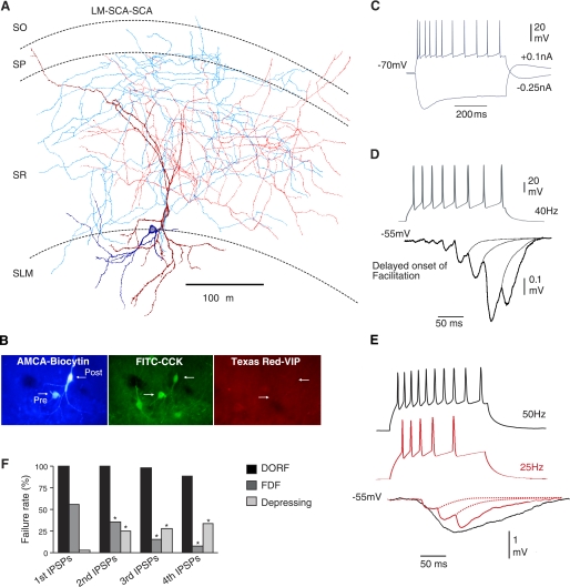
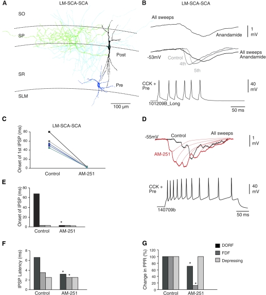
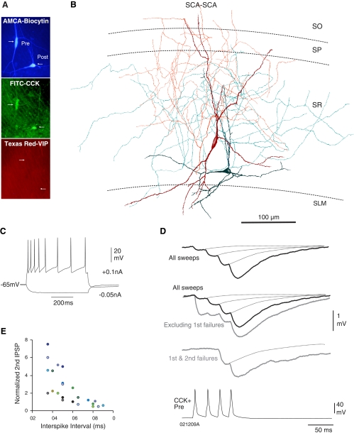
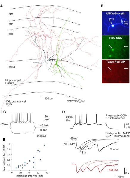
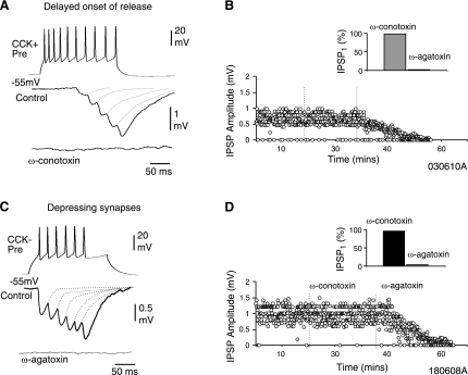
One of the critical factors in determining network behavior of neurons is the influence of local circuit connections among interneurons. The short-term synaptic plasticity and the subtype of presynaptic calcium channels used at local circuit connections of inhibitory interneurons in CA1 were investigated using dual whole-cell recordings combined with biocytin and double immunofluorescence labeling in acute slices of P18- to 21-day-old rat stratum radiatum (SR) and stratum lacunosum molecular (SLM). Two forms of temporally distinct synaptic facilitation were observed among interneuron connections involving presynaptic cholecystokinin (CCK)-positive cells in SR, frequency-dependent facilitation, and a delayed onset of release (45-80 ms) with subsequent facilitation (DORF). Inhibition at both these synapses was under tonic cannabinoid-type 1 (CB1) receptor activity. DORF synapses did not display conventional release-dependent properties; however, blocking CB1 receptors with antagonist AM-251 (10 μM) altered the synaptic transmission to frequency-dependent depression with a fast onset of release (2-4 ms). Presynaptic CCK-negative interneurons in SLM elicited inhibitory postsynaptic potentials (IPSPs) insensitive to CB1 receptor pharmacology displayed frequency-dependent depression. Release of GABA at facilitating synapses was solely mediated via N-type presynaptic calcium channels, whereas depressing synapses utilized P/Q-type channels. These data reveal two distinct models of neurotransmitter release patterns among interneuron circuits that correlate with the subtype of presynaptic calcium channel. These data suggest that endocannabinoids act via CB1 receptors to selectively modulate N-type calcium channels to alter signal transmission.


Figures

Fig. 1.
Fig. 1.
Temporally distinct synaptic facilitation that is delayed on the onset of release. A: reconstruction of a synaptically connected stratum lacunosum moleculare (SLM) interneuron, lacunosum moleculare-Shaffer collateral associated cell (LM-SCA; blue-soma, light blue-axon), and a stratum radiatum (SR), Shaffer collateral associated cell (SCA; deep red- dendrites, red-axon). B: double immunoflorescence labeling revealed the two recorded cells filled with biocytin (AMCA) immunopositive for CCK (FITC) and immunonegative for VIP (Texas red). C: intrinsic membrane properties of the presynaptic LM-SCA cell. In response to hyperpolarizing current injection this interneuron displayed pronounced sag, indicative of Ih current. In response to depolarizing current fast but adapting firing patterns were displayed. D: IPSPs elicited by LM-SCA cells displayed a delayed onset of synaptic facilitation (DORF). IPSPs were usually observed after third to fifth presynaptic spike in a train. E: another synaptic connection between a LM-SCA cell and SCA cell; the onset of first IPSP did not appear earlier in the train when increasing the firing frequency of the presynaptic interneuron from 25 Hz to 50 Hz. At higher presynaptic firing frequencies the IPSPs summated more readily, shown by the black trace. F: comparison of average failure rates of 1st, 2nd, 3rd and 4th IPSPs in control at DORF, frequency dependent facilitating (FDF), and depressing synapses studied. SP, stratum pyramidale, SO, stratum oriens.
Fig. 2.
Fig. 2.
Temporary release-independence via CB1 receptors at synapses displaying DORF. A: reconstruction of a LM-SCA and a basket cell (black soma, green axon). B: bath application of 14 μM anandamide (CB receptor agonist) enhanced the facilitation. C: the change in the onset of release of GABA in control and in AM-251; data for individual pairs are shown (n = 5). D: the delayed release and suppression of inhibition was blocked by 10 μM CB1 receptor antagonist, AM-251, changing the synaptic efficacy and the onset of the IPSPs, which appeared earlier during the train of action potentials elicited. E and F: bar graphs comparing the onset of first IPSPs during a train of presynaptic action potentials and IPSP latency at DORF, FDF, and depressing synapse in control and after bath application of AM-251. Onset of the first IPSP seem to be delayed at connections between LM-SCA and SCA interneurons. G: comparison of change in paired-pulse ratios (PPRs) in control and AM-251. *Significantly different than control first IPSPs.
Fig. 3.
Fig. 3.
Synaptic facilitation between 2 CCK-positive, SCA interneurons is release dependent. A: immunofluorescence labeling showing both recorded cells as CCK positive and VIP negative. B: reconstruction of the biocytin-labeled cells shown in A, which displayed synaptic facilitation shown in D. C: firing properties of the presynaptic SCA interneuron (shown in red below) recorded in response to −0.05 and 0.1 nA current injection. SCA cells typically displayed adapting properties (see Table 1). D: averaged responses to trains of presynaptic action potentials (black traces include all sweeps in average; gray traces include specific subsets of data). These connections typically displayed synaptic facilitation, which was greater after excluding first and second IPSP failures or only first IPSP failures. Excluding first IPSP failures resulted in larger first and smaller second IPSPs (gray traces). E: graph to illustrate the recovery of second IPSP from synaptic facilitation at longer interspike intervals (each color represents an individual pair of connection, n = 12).
Fig. 4.
Fig. 4.
CCK-negative, long-range quadrilaminar cells display synaptic depression. A: reconstruction of a synaptically connected CCK-negative quadrilaminar cell (green soma, red axon) to a LM-SCA cell (gray). B: double immunoflorescence labeling reveals the 2 recorded cells filled with biocytin (AMCA) immunonegative for VIP (Texas red) and only the postsynaptic cell was immunopositive for CCK (FITC). C: intrinsic membrane properties of the presynaptic back projecting cell shown in A and B. D: this synapse displayed release of neurotransmitter at short latencies of 2.5 ms with precision. IPSPs elicited by a train of action potentials displayed synaptic depression. The gray traces are superimposed averages of second IPSPs at different spike intervals; the shorter the spike interval, the stronger the depression (illustrated by smaller IPSPs). These IPSPs were insensitive to AM-251 (red trace). E: graph to illustrate the recovery from synaptic depression at longer inter spike intervals (each color represents an individual pair, n = 8).
Fig. 5.
Fig. 5.
Different calcium channels mediate synaptic transmission at depressing, CB1-insensitive, and facilitating, CB1-sensitive synapses. A: ω-conotoxin GVIa (1 μM) blocks transmission at CCK-positive presynaptic interneurons that demonstrate a delayed onset of release and synaptic facilitation, suggesting the involvement of N-type calcium channels. Facilitating synapses sensitive to CB1 receptor pharmacology were not sensitive to ω-agatoxin Iva. B: plot of peak amplitudes of second IPSPs during an experiment in control and in bath application of calcium channel blockers. B, Inset: summary of normalized unitary IPSP amplitudes in ω-agatoxin and ω-conotoxin (n = 4). C: presynaptic CCK-negative interneurons elicited depressing IPSPs and was insensitive to ω-conotoxin GVIa (1 μM), but transmission was blocked by ω-agatoxin IVa (0.5 μM), suggesting the involvement of P/Q-type calcium channels. D: plot of peak amplitudes of first IPSPs during an experiment in control and in bath application of calcium channel blockers. D, Inset: summary of normalized unitary IPSP amplitudes in ω-conotoxin and ω-agatoxin (n = 3).
Fig. 6.
Fig. 6.
Three types of short-term synaptic plasticity at local circuit interneuron connections in CA1. CCK-positive interneurons in SR (where glutamatergic input from CA3 enters) receive facilitating IPSPs, which are temporally distinct (FDF and DORF), mediated by N-type calcium channels and under the modulation via CB1 receptors. Synaptic depression is observed in SP and SLM (mediated by P/Q-type calcium channel and insensitive to CB1 receptor pharmacology) where excitatory input from entorhinal cortex and the thalamus are received. This localized short-term plasticity among interneuron synapses probably plays important roles in synaptic integration while rapidly shunting some inhibitory signals; other signals are allowed a wider time window for synaptic integration. PV, parvalbumin.

References

    1. Ali AB. Presynaptic inhibition of GABAA receptor-mediated unitary IPSPs by cannabinoid receptors at synapses between CCK-positive interneurons in rat hippocampus. J Neurophysiol 98: 861–869, 2007 - PubMed
    1. Ali AB, Bannister AP, Thomson AM. IPSPs elicited in CA1 pyramidal cells by putative basket cells in slices of adult rat hippocampus. Eur J Neurosci 11: 1741–1753, 1999 - PubMed
    1. Ali AB, Deuchars J, Pawelzik H, Thomson AM. CA1 pyramid to basket and bistratified cells EPSPs: dual intracellular recordings in rat hippocampal slices. J Physiol 507.1: 201–217, 1998 - PMC - PubMed
    1. Ali AB, Nelson C. Distinct Ca2+ channels mediate transmitter release at excitatory synapses displaying different dynamic properties in rat neocortex. Cereb Cortex 6: 386–393, 2006 - PubMed
    1. Ali AB, Todorova M. Asynchronous release of GABA via tonic cannabinoid receptor activation at identified interneuron synapses in rat CA1. Eur J Neurosci 31: 3–12, 2010 - PubMed

Publication types

Substances