. 2010 Dec 21;107(51):22207-12.
doi: 10.1073/pnas.1015954107. Epub 2010 Dec 1.

Elements between the IgH variable (V) and diversity (D) clusters influence antisense transcription and lineage-specific V(D)J recombination

Affiliations

Elements between the IgH variable (V) and diversity (D) clusters influence antisense transcription and lineage-specific V(D)J recombination

Cosmas C Giallourakis et al. Proc Natl Acad Sci U S A. .

Abstract

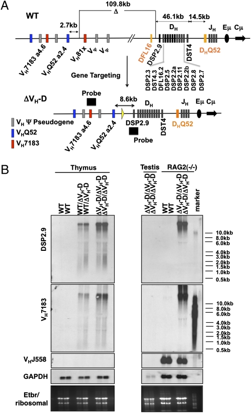
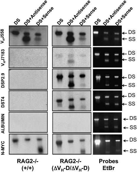
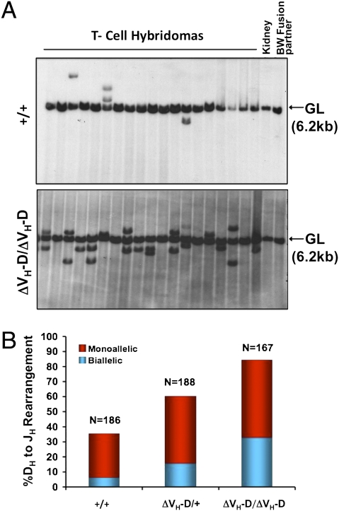
Ig and T-cell receptor (TCR) variable-region gene exons are assembled from component variable (V), diversity (D) and joining (J) gene segments during early B and T cell development. The RAG1/2 endonuclease initiates V(D)J recombination by introducing DNA double-strand breaks at borders of the germ-line segments. In mice, the Ig heavy-chain (IgH) locus contains, from 5' to 3', several hundred V(H) gene segments, 13 D segments, and 4 J(H) segments within a several megabase region. In developing B cells, IgH variable-region exon assembly is ordered with D to J(H) rearrangement occurring on both alleles before appendage of a V(H) segment. Also, IgH V(H) to DJ(H) rearrangement does not occur in T cells, even though DJ(H) rearrangements occur at low levels. In these contexts, V(D)J recombination is controlled by modulating substrate gene segment accessibility to RAG1/2 activity. To elucidate control elements, we deleted the 100-kb intergenic region that separates the V(H) and D clusters (generating ΔV(H)-D alleles). In both B and T cells, ΔV(H)-D alleles initiated high-level antisense and, at lower levels, sense transcription from within the downstream D cluster, with antisense transcripts extending into proximal V(H) segments. In developing T lymphocytes, activated germ-line antisense transcription was accompanied by markedly increased IgH D-to-J(H) rearrangement and substantial V(H) to DJ(H) rearrangement of proximal IgH V(H) segments. Thus, the V(H)-D intergenic region, and likely elements within it, can influence silencing of sense and antisense germ-line transcription from the IgH D cluster and thereby influence targeting of V(D)J recombination.


Conflict of interest statement

The authors declare no conflict of interest.

Figures

Fig. 1.
Fig. 1.
Deletion of VH to DH intergenic region leads to generation of long VH- and D-containing transcripts. (A) Schematic diagram of proximal WT murine 129sVe IgH locus before (Upper) and after gene targeting to delete the 109-kb intergenic region (Lower). The order of the D segments is indicated. (B) Northern analyses of total RNA from WT (+/+), ΔVH-D/+, and ΔVH-D/ΔVH-D thymocytes (Left) as well as total RNA from RAG2−/−(+/+) and RAG2−/−(ΔVH-D/ΔVH-D) A-MuLV-transformed pro-B lines after probing with (from top to bottom): a 1-kb fragment encompassing DSP2.9 (see A); a 511-bp fragment derived from the germ-line VH7183.a4.6 segment (see A); a 631-bp probe derived from the germ line corresponding to the VHJ558.29 segment; and a GAPDH cDNA. Ribosomal RNA loading is shown at the bottom as additional loading control.
Fig. 2.
Fig. 2.
Deletion of VH to DH intergenic region leads increased germ-line transcription. Nuclear run-ons were conducted with nuclei from RAG2−/− (+/+) and RAG2−/−(ΔVH-D/ΔVH-D) A-MuLV-transformed pro-B lines. 32P-UTP–labeled nascent transcripts were hybridized to a series of double-stranded restriction endonuclease-generated DNA fragments (named on x axes) that were separated by electrophoresis and immobilized on Southern membranes. (A) The location of nuclear run-on probes (A through S) within the WT (Upper) and ΔVH-D (Lower) IgH alleles is shown. (B) Schematic diagram of location of probe fragments in gels shown in C and D. Open squares depict backbone vector sequences, and filled squares depict IgH fragments/probes. (C and D) (Upper) Ethidium bromide–stained gel of Southern membrane. (Lower) Same membrane exposed after probing with labeled transcripts from RAG2−/− (+/+) (Left) and RAG2−/−(ΔVH-D/ΔVH-D) (Right) A-MuLV-transformed pro-B lines. Results are representative of three independent experiments with different lines of the indicated genotype. See SI Materials and Methods for details.
Fig. 3.
Fig. 3.
Deletion of VH to DH intergenic region leads to antisense transcription of adjacent regions. Nuclear run-ons were conducted with nuclei from RAG2−/− (+/+) and RAG2−/−(ΔVH-D/ΔVH-D) A-MuLV-transformed pro-B lines. 32P-UTP–labeled nascent transcripts were hybridized to indicated double strand (DS), antisense, and sense DNA fragments from the IgH and control loci as indicated (see text and SI Materials and Methods for further details). (Left and Center) Run-on results. (Right) Loading as revealed by ethidium bromide staining. SS, single strand.
Fig. 4.
Fig. 4.
VH to DH intergenic deletion up-regulates DH to JH recombination in thymocytes. (A) Two sets of independent splenic T-cell hybridomas were obtained from WT (n = 186), ΔVH-D/+ (n = 188), and ΔVH-D/ΔVH-D (n = 167) mice. DNA was digested with StuI, and Southern blot analysis was performed with an EcoRI/HindIII fragment probe downstream of JH4. The germ-line JH locus yields a 6.2-kb band (GL), and DH to JH rearrangements are scored as rearranged bands with a size distinct from the germ-line band. DNA from the fusion partner BW5147 and kidney DNA is shown as controls. A representative set of analyses is shown. (B) Percentage of cells containing monoallelic (red) and biallelic (blue) JH rearrangements as determined by either one or two non-germ-line bands in analyses of A. The average of two independent experiments is shown.
Fig. 5.
Fig. 5.
Deletion of the VH to DH intergenic region leads to VH to DJH recombination in thymocytes. (A) PCR assay for VH to DJH for indicated gene families (Right) in DNA from sorted DP-T cells. Size of rearrangements to various J segments is indicated on Left. Analysis of Igκ rearrangement (VJκ) controlled for potential B cell contamination. (B) The level of VH to DJH rearrangements in the same panel of hybridomas shown in Fig. 4 was determined by a combination of hybridization to a JH probe, a probe between VH and DH segments, plus PCR for proximal VH to DJH rearrangements in each hybridoma (see SI Materials and Methods for details).

Similar articles

Cited by

References

    1. Schatz DG. V(D)J recombination. Immunol Rev. 2004;200:5–11. - PubMed
    1. Jung D, Giallourakis C, Mostoslavsky R, Alt FW. Mechanism and control of V(D)J recombination at the immunoglobulin heavy chain locus. Annu Rev Immunol. 2006;24:541–570. - PubMed
    1. Yancopoulos GD, Blackwell TK, Suh H, Hood L, Alt FW. Introduced T cell receptor variable region gene segments recombine in pre-B cells: Evidence that B and T cells use a common recombinase. Cell. 1986;44:251–259. - PubMed
    1. Schatz DG, Baltimore D. Uncovering the V(D)J recombinase. Cell. 2004;116:S103–S106. - PubMed
    1. Fugmann SD, Lee AI, Shockett PE, Villey IJ, Schatz DG. The RAG proteins and V(D)J recombination: Complexes, ends, and transposition. Annu Rev Immunol. 2000;18:495–527. - PubMed

Publication types

MeSH terms