. 2010 Dec 1;2(60):60ra89.
doi: 10.1126/scitranslmed.3001380.

Replacing the enzyme alpha-L-iduronidase at birth ameliorates symptoms in the brain and periphery of dogs with mucopolysaccharidosis type I

Affiliations

Replacing the enzyme alpha-L-iduronidase at birth ameliorates symptoms in the brain and periphery of dogs with mucopolysaccharidosis type I

Ashley D Dierenfeld et al. Sci Transl Med. .

Abstract

Mucopolysaccharidosis type I (MPS I) is a lysosomal storage disease caused by loss of activity of α-l-iduronidase and attendant accumulation of the glycosaminoglycans dermatan sulfate and heparan sulfate. Current treatments are suboptimal and do not address residual disease including corneal clouding, skeletal deformities, valvular heart disease, and cognitive impairment. We treated neonatal dogs with MPS I with intravenous recombinant α-l-iduronidase replacement therapy at the conventional 0.58 mg/kg or a higher 1.57 mg/kg weekly dose for 56 to 81 weeks. In contrast to previous results in animals and patients treated at a later age, the dogs failed to mount an antibody response to enzyme therapy, consistent with the induction of immune tolerance in neonates. The higher dose of enzyme led to complete normalization of lysosomal storage in the liver, spleen, lung, kidney, synovium, and myocardium, as well as in the hard-to-treat mitral valve. Cardiac biochemistry and function were restored, and there were improvements in skeletal disease as shown by clinical and radiographic assessments. Glycosaminoglycan levels in the brain were normalized after intravenous enzyme therapy, in the presence or absence of intrathecal administration of recombinant α-l-iduronidase. Histopathological evidence of glycosaminoglycan storage in the brain was ameliorated with the higher-dose intravenous therapy and was further improved by combining intravenous and intrathecal therapy. These findings argue that neonatal testing and early treatment of patients with MPS I may more effectively treat this disease.


Conflict of interest statement

Competing interests: The authors have no additional relationships to disclose. The Los Angeles Biomedical Research Institute at Harbor-UCLA and the Harbor-UCLA Department of Pediatrics hold patent interests in rhIDU.

Figures

Fig. 1
Fig. 1
Enrollment and induction of immune tolerance to IDU. (A) Study enrollment timeline for animals receiving 0.58 mg/kg weekly rhIDU (n=8), indicating age at first rhIDU dose (days) and age at sacrifice (weeks). Animals receiving concomitant intrathecal (IT) ERT are indicated with *. IT ERT was administered at a dose of 0.058 mg/kg (maximum 1 mg) at three-month intervals. (B) Enrollment timeline for dogs treated with 1.57 mg/kg weekly IV rhIDU (n=4). (C) Serum anti-iduronidase IgG antibodies measured by ELISA, depicting pretreatment and final levels and expressed as OD units per μl undiluted serum on a semi-log scale. Tolerance is defined as an IgG titer of less than 20 OD units/μl. Positive non-tolerant control shown for comparison (12, 13, 21).
Fig. 2
Fig. 2
Glycosaminoglycan (GAG) storage in tissues in dogs treated with IV rhIDU from birth. (A–F) Glycosaminoglycan concentrations measured by Alcian blue dye-binding assay in kidney medulla, kidney cortex, liver, spleen, lung and myocardium of normal dogs, MPS I dogs treated neonatally with 1.57 or 0.58 mg/kg weekly IV rhIDU, and untreated MPS I dogs. Each dot represents one animal. Means and standard deviations were compared by ANOVA. Pairwise post-hoc analysis involved the Tukey-Kramer adjustment and p values are indicated at the top of each graph.
Fig. 3
Fig. 3
Effects of IV rhIDU administration beginning at birth on cardiac disease in MPS I dogs. (A) The GAG storage in the canine mitral valve in normal dogs, MPS I dogs receiving 1.57 mg/kg or 0.58mg/kg weekly IV rhIDU, and untreated MPS I dogs. P-values show comparison of treatment group means to the mean of untreated MPS I dogs (ANOVA with Tukey-Kramer post-hoc analysis). (B) Average thickness (mm) of the anterior mitral leaflet measured by 2-D echocardiography in diastole. P-values show comparison of treatment group means to the mean of untreated MPS I dogs. Each reading is the average of 3 to 15 measurements per dog. (C-E) Images of mitral valve by echocardiography in (C) a normal dog, (D) a 0.58 mg/kg treated MPS I dog, and (E) an untreated MPS I dog. A normal electrocardiogram trace is seen at the bottom of each panel. (F–I) Histopathology of the heart valve (right atrioventricular valve). Black bar indicates 10 μm. (F) Normal, (G) MPS I affected animal treated with 1.57 mg/kg weekly IV, (H) MPS I affected animal treated with 0.58 mg/kg weekly IV, with mild residual lysosomal (GAG) storage indicated by arrowheads, and (I) an untreated affected MPS I dog (lysosomal storage indicated by arrowheads).
Fig. 4
Fig. 4
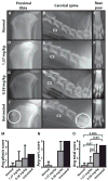
Effects of IV rhIDU from birth on skeletal abnormalities in MPS I dogs (A–I). Treated dogs averaged 12.8 ± 0.2 months (1.57 mg/kg), or 17.1± 1.0 months (0.58 mg/kg) of age, affected dogs 18.1 ± 1.0 months, and normal dogs 18.4 ± 3.2 months. (AC) Radiographs of normal, (D–F) 1.57 mg/kg treated and (G–I) 0.58 mg/kg treated MPS I affected canines, and (J–L) untreated MPS I affected canines. C3 in (B, E, H, K), the third cervical vertebra. A physeal remnant/open physis (circled) is seen in an untreated 17 month old animal (J). (K) Severe narrowing of the intervertebral disc space (circled) in an untreated, affected dog and moderate intervertebral space narrowing in a 0.58 mg/kg weekly treated dog (H), not seen in (B), a normal dog, or (E), a 1.57 mg/kg/wk treated dog. Also evident in (K) is C2-C3 narrowing of the intervertebral space. (L) Toe splaying seen in untreated animal, but not seen in normal or treated dogs (C, F, and I). (M–O) Scoring of skeletal disease indicating average group (M) physis, (N) vertebra, and (O) total scores (see scoring in materials and methods). The two untreated animals depicted in this graph received the same score, resulting in a lack of error bars. Pairwise P values are indicated in the total score bar graph.
Fig. 5
Fig. 5
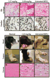
Effects of IV rhIDU from birth on rib periosteum, joints, skull morphology, joint laxity, and cornea. (A–C) Histopathology of rib periosteum. (D–F) Histopathology of synovium around patella. Untreated affected MPS I (C and F) shows storage of GAGs in both of these tissues, as evidenced by the distended cells. Reduced storage is shown in treated animals (A–B, D–E), with no storage evident in the 1.57 mg/kg treated animals (A, D). (G–I) Skull morphology and (J–L) carpal laxity of normal dogs (G and J), MPS I affected animals receiving 1.57 mg/kg (H and K), and untreated MPS I affected dogs (I and L). Signs of skull abnormalities include a dome-shaped head, a pronounced stop (depressed nasal bridge) and an upturned muzzle (I). (M–O) Hematoxylin and eosin staining of the cornea from a normal dog (M), a dog treated with 1.57 mg/kg (N), and an untreated MPS I dog (O). Intracellular storage in the corneal stroma appears pale and foamy.
Fig. 6
Fig. 6
Effects of IV and IT rhIDU on the brain in MPS I dogs. (A) Magnetic resonance imaging of the brain (T1-weighted with contrast enhancement) in untreated MPS I dog, showing meningiomas (arrows, A). (B–C) Low power (10x, B, Bar = 200 μm) and high-power (40x, C, bar = 20 μm) magnification of a fibrous meningioma in the same untreated MPS I dog. Foamy interstitial cells are indicated by arrows. Lymphoplasmacytic aggregates within the mass are indicated by an arrowhead. (D–F) Toluidine blue staining of cortical neurons in MPS I dogs. Untreated (D), 1.57 mg/kg IV treated (E) and IV/IT treated (F), with lipid granules indicated by arrows. (G–H) Histograms showing glycosaminoglycan concentrations (G) and iduronidase activity (H) in brain of normal, treated MPS I dogs, and untreated MPS I dogs. For iduronidase activity (H), a log10 scale is used for the y-axis. Samples from each of six coronal sections were averaged for each dog, and these means were then averaged for all dogs in the group. ANOVA with post-hoc Tukey-Kramer analysis was used to determine p-values.

References

    1. Bunge S, Clements PR, Byers S, Kleijer WJ, Brooks DA, Hopwood JJ. Genotype-phenotype correlations in mucopolysaccharidosis type I using enzyme kinetics, immunoquantification and in vitro turnover studies. Biochim Biophys Acta. 1998;1407:249–256. - PubMed
    1. Neufeld EF, Muenzer J. In: The Metabolic and Molecular Bases of Inherited Disease. Scriver CR, Beaudet AL, Sly WS, Valle D, editors. McGraw-Hill, Health Professions Division; New York: 2001. pp. 3421–3452.
    1. Soper BW, Duffy TM, Lessard MD, Jude CD, Schuldt AJ, Vogler CA, Levy B, Barker JE. Transplanted ER-MP12hi20-58med/hi myeloid progenitors produce resident macrophages from marrow that are therapeutic for lysosomal storage disease. Blood Cells Mol Dis. 2004;32:199–213. - PubMed
    1. Whitley CB, Belani KG, Chang PN, Summers CG, Blazar BR, Tsai MY, Latchaw RE, Ramsay NK, Kersey JH. Long-term outcome of Hurler syndrome following bone marrow transplantation. Am J Med Genet. 1993;46:209–218. - PubMed
    1. Pastores GM, Arn P, Beck M, Clarke JT, Guffon N, Kaplan P, Muenzer J, Norato DY, Shapiro E, Thomas J, Viskochil D, Wraith JE. The MPS I registry: design, methodology, and early findings of a global disease registry for monitoring patients with Mucopolysaccharidosis Type I. Mol Genet Metab. 2007;91:37–47. - PubMed

Publication types

LinkOut - more resources