

Noninvasive imaging of pancreatic islet inflammation in type 1A diabetes patients
- PMID: 21123946
- PMCID: PMC3007157
- DOI: 10.1172/JCI44339
Noninvasive imaging of pancreatic islet inflammation in type 1A diabetes patients
Abstract
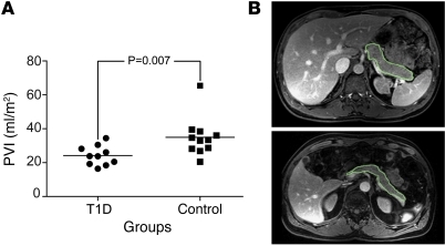
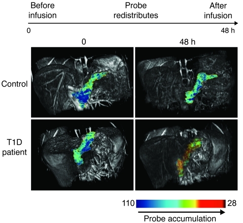
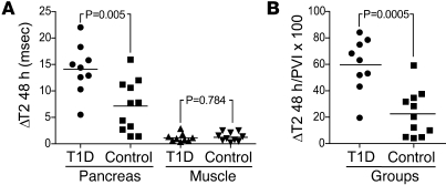
Type 1A diabetes (T1D) is an autoimmune disease characterized by leukocyte infiltration of the pancreatic islets of Langerhans. A major impediment to advances in understanding, preventing, and curing T1D has been the inability to "see" the disease initiate, progress, or regress, especially during the occult phase. Here, we report the development of a noninvasive method to visualize T1D at the target organ level in patients with active insulitis. Specifically, we visualized islet inflammation, manifest by microvascular changes and monocyte/macrophage recruitment and activation, using magnetic resonance imaging of magnetic nanoparticles (MNPs). As a proof of principle for this approach, imaging of infused ferumoxtran-10 nanoparticles permitted effective visualization of the pancreas and distinction of recent-onset diabetes patients from nondiabetic controls. The observation that MNPs accumulate in the pancreas of T1D patients opens the door to exploiting this noninvasive imaging method to follow T1D progression and monitoring the ability of immunomodulatory agents to clear insulitis.
Figures
References
-
- Foulis AK, Liddle CN, Farquharson MA, Richmond JA, Weir RS. The histopathology of the pancreas in type 1 (insulin-dependent) diabetes mellitus: a 25-year review of deaths in patients under 20 years of age in the United Kingdom. Diabetologia. 1986;29(5):267–274. doi: 10.1007/BF00452061. - DOI - PubMed
-
- Itoh N, et al. Mononuclear cell infiltration and its relation to the expression of major histocompatibility complex antigens and adhesion moleucles in pancreas biospy speciments from newly diagnosed insulin-dependent diabetes mellitus patients. J Clin Invest. 1993;92(5):2313–2322. doi: 10.1172/JCI116835. - DOI - PMC - PubMed
-
- Papaccio G. Insulitis and islet microvasculature in type 1 diabetes. Histol Histopathol. 1993;8(4):751–759. - PubMed
Publication types
MeSH terms
Substances
Grants and funding
LinkOut - more resources
Full Text Sources
Other Literature Sources
Medical
Research Materials
