Case Reports
. 2011 Jan;121(1):113-9.
doi: 10.1172/JCI44073. Epub 2010 Dec 1.

Adult combined GH, prolactin, and TSH deficiency associated with circulating PIT-1 antibody in humans

Affiliations
Case Reports

Adult combined GH, prolactin, and TSH deficiency associated with circulating PIT-1 antibody in humans

Masaaki Yamamoto et al. J Clin Invest. 2011 Jan.

Abstract

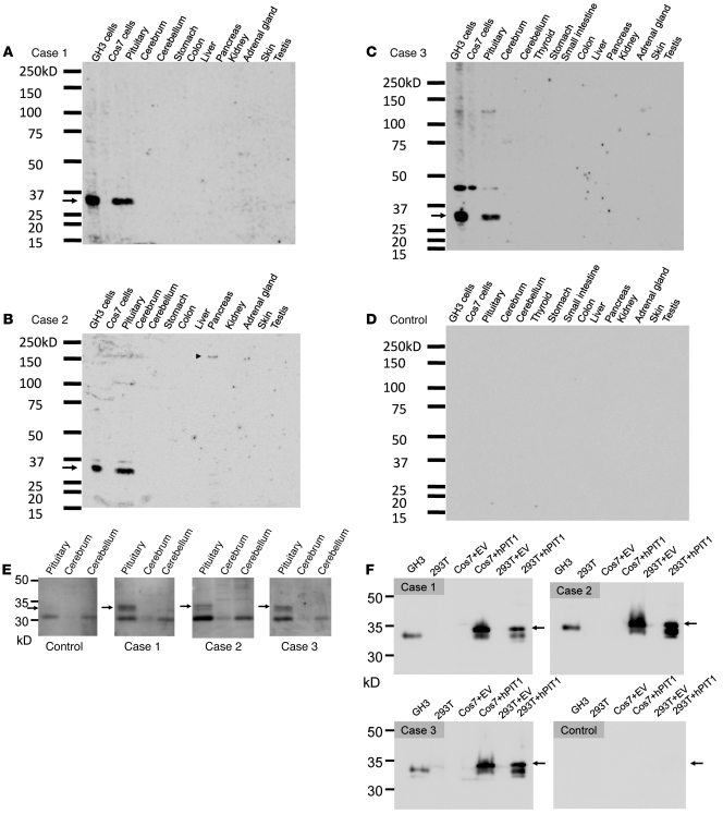
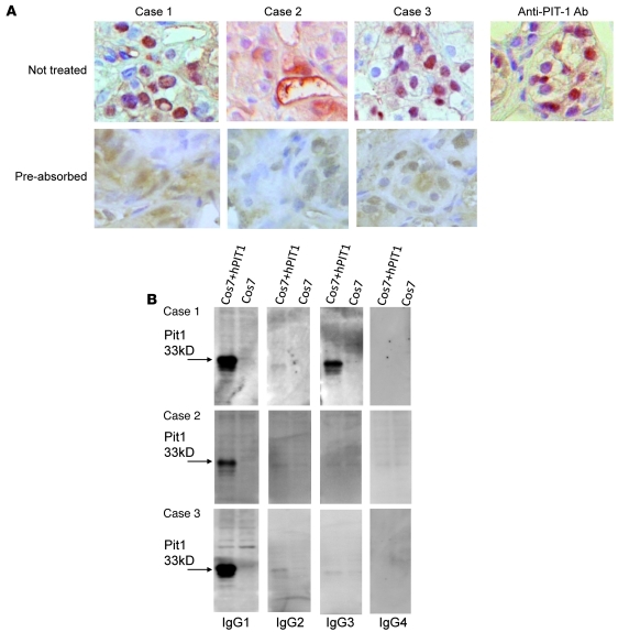
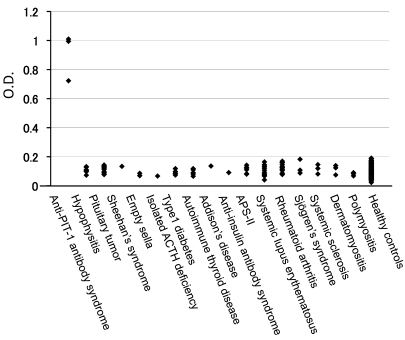
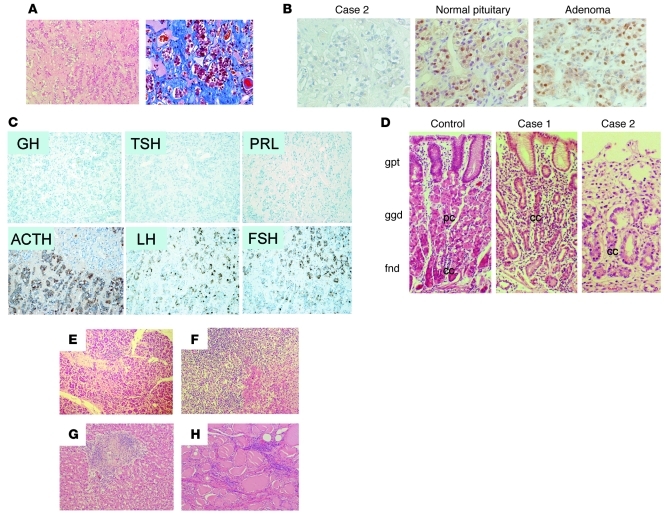
The pituitary-specific transcriptional factor-1 (PIT-1, also known as POU1F1), is an essential factor for multiple hormone-secreting cell types. A genetic defect in the PIT-1 gene results in congenital growth hormone (GH), prolactin (PRL), and thyroid-stimulating hormone (TSH) deficiency. Here, we investigated 3 cases of adult-onset combined GH, PRL, and TSH deficiencies and found that the endocrinological phenotype in each was linked to autoimmunity directed against the PIT-1 protein. We detected anti-PIT-1 antibody along with various autoantibodies in the patients' sera. An ELISA-based screening revealed that this antibody was highly specific to the disease and absent in control subjects. Immunohistochemical analysis revealed that PIT-1-, GH-, PRL-, and TSH-positive cells were absent in the pituitary of patient 2, who also had a range of autoimmune endocrinopathies. These clinical manifestations were compatible with the definition of autoimmune polyendocrine syndrome (APS). However, the main manifestations of APS-I--hypoparathyroidism and Candida infection--were not observed and the pituitary abnormalities were obviously different from the hypophysitis associated with APS. These data suggest that these patients define a unique "anti-PIT-1 antibody syndrome," related to APS.


Figures

Figure 1
Figure 1. Immunoblotting analysis of mouse tissues and cell lysates.
The patients’ serum was used as a primary antibody (1:1000) to detect the autoantibody. (AC) The patients’ sera specifically recognized a 33-kDa protein in the lysates from the pituitary and GH3 cells (arrow). (B) The serum of patient 2 recognizes a 150-kDa protein in the pancreas (arrowhead), but the size of this protein does not correspond to that of insulin (58 kDa) or GAD (65 kDa). (C) The serum of patient 3 recognized approximately 45 kDa protein as a nonspecific band in addition to the 33-kDa protein PIT-1. (D) Representative results of the healthy control subjects are shown. Neither the sera of 10 healthy control subjects nor those of 8 patients with pituitary adenoma and 6 patients with hypophysitis recognized the 33-kDa protein. (E) The patients’ sera detected the 33-kDa protein in the lysate from human pituitary. (F) The patients’ sera detected the PIT-1 protein in the lysates from GH3, hPIT-1–expressing Cos7, and 293T cells (arrows).
Figure 2
Figure 2. Patients’ sera detected natural form of PIT-1 by immunohistochemistry, and the antibodies were of IgG1 and IgG3 subtypes.
(A) Immunohistochemical analysis revealed that patients’ sera, as well as anti–PIT-1 antibody, recognize human anterior pituitary cells in the normal pituitary tissue. Original magnification, ×600. Preabsorption of the sera with rhPIT-1 substantially reduced the signal, indicating the specificity for PIT-1. (B) The anti–PIT-1 antibody belongs to the IgG1 and IgG3 fractions in case 1 and to the IgG1 fraction in cases 2 and 3 (B and Supplemental Figure 3E).
Figure 3
Figure 3. An anti–PIT-1 antibody–specific ELISA demonstrated that the anti–PIT-1 antibody is highly specific to these patients.
This antibody was not detected in control subjects or patients with other endocrine and autoimmune diseases, including APS-II.
Figure 4
Figure 4. Histological analysis revealed the defect in PIT-1–positive cells and multiple endocrine organopathy.
(A) Microscopic analysis of H&E-stained sections of the pituitary tissue from patient 2. The number of adenohypophyseal cells is markedly reduced. The reduction is accompanied by infiltration of lymphocytes and plasma cells (left panel) and mild fibrosis in the stromal tissue (right panel: Azan staining). (B) Immunohistochemical analysis using an anti–PIT-1 antibody. In contrast to the normal pituitary or pituitary adenoma, no PIT-1–positive cells are observed in the pituitary in the case of patient 2. (C) Immunohistochemistry of the pituitary in the case of patient 2. GH-, PRL-, and TSH-positive cells are absent, despite the presence of ACTH-, LH-, and FSH-positive cells. (D) Histological analysis of the gastric mucosa from control and patients 1 and 2. In contrast to the control, no parietal cells were detected in patients 1 and 2. gpt, gastric pit; ggd, gastric glands; fnd, fundic glands; pc, parietal cells; cc, chief cells. Histological analysis of tissue of the pancreas (E), adrenal gland (F), liver (G), and thyroid (H) from patient 2 revealed inflammatory infiltration and disrupted tissue structure. Original magnification, ×100 (A, C); ×400 (B); ×200 (D); ×100 (EH).

References

    1. Ingraham HA, et al. A tissue-specific transcription factor containing a homeodomain specifies a pituitary phenotype. Cell. 1988;55(3):519–529. doi: 10.1016/0092-8674(88)90038-4. - DOI - PubMed
    1. Cohen LE, Wondisford FE, Radovick S. Role of Pit-1 in the gene expression of growth hormone, prolactin, and thyrotropin. Endocrinol Metab Clin North Am. 1996;25(3):523–540. doi: 10.1016/S0889-8529(05)70339-X. - DOI - PubMed
    1. Pfaffle RW, et al. Mutation of the POU-specific domain of Pit-1 and hypopituitarism without pituitary hypoplasia. Science. 1992;257(5073):1118–1121. doi: 10.1126/science.257.5073.1118. - DOI - PubMed
    1. Michels AW, Gottlieb PA. Autoimmune polyglandular syndromes. Nat Rev Endocrinol. 2010;6(5):270–277. doi: 10.1038/nrendo.2010.40. - DOI - PubMed
    1. Finnish-German APECED Consortium. An autoimmune disease, APECED, caused by mutations in a novel gene featuring two PHD-type zinc-finger domains. Nat Genet. 1997;17(4):399–403. doi: 10.1038/ng1297-399. - DOI - PubMed

Publication types

MeSH terms