

Inactivation of Bmp4 from the Tbx1 expression domain causes abnormal pharyngeal arch artery and cardiac outflow tract remodeling
- PMID: 21123999
- PMCID: PMC3124451
- DOI: 10.1159/000321170
Inactivation of Bmp4 from the Tbx1 expression domain causes abnormal pharyngeal arch artery and cardiac outflow tract remodeling
Abstract
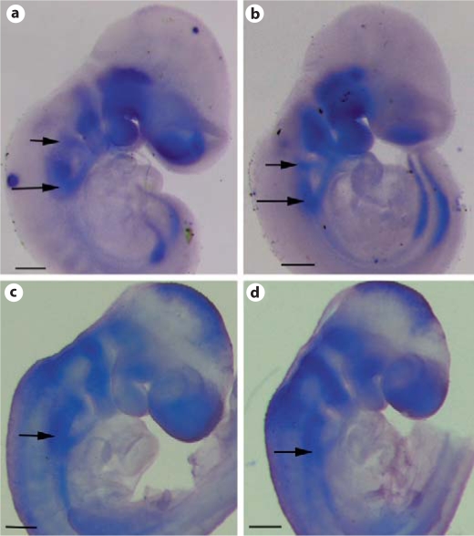
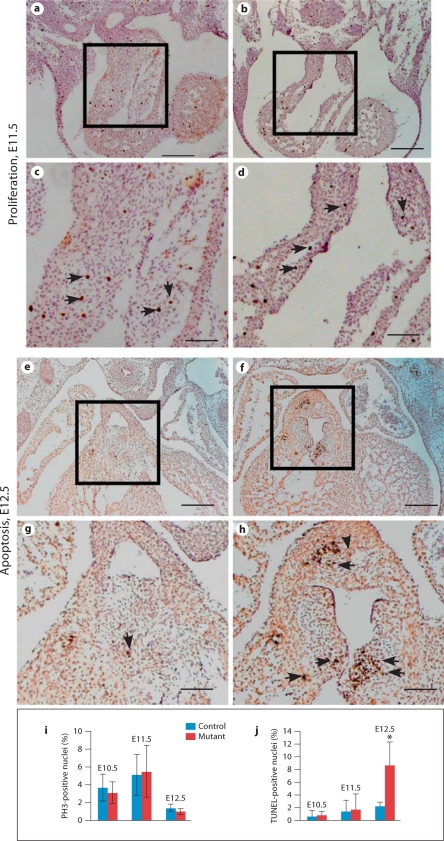
Maldevelopment of outflow tract and aortic arch arteries is among the most common forms of human congenital heart diseases. Both Bmp4 and Tbx1 are known to play critical roles during cardiovascular development. Expression of these two genes partially overlaps in pharyngeal arch areas in mouse embryos. In this study, we applied a conditional gene inactivation approach to test the hypothesis that Bmp4 expressed from the Tbx1 expression domain plays a critical role for normal development of outflow tract and pharyngeal arch arteries. We showed that inactivation of Bmp4 from Tbx1-expressing cells leads to the spectrum of deformities resembling the cardiovascular defects observed in human DiGeorge syndrome patients. Inactivation of Bmp4 from the Tbx1 expression domain did not cause patterning defects, but affected remodeling of outflow tract and pharyngeal arch arteries. Our further examination revealed that Bmp4 is required for normal recruitment/differentiation of smooth muscle cells surrounding the PAA4 and survival of outflow tract cushion mesenchymal cells.
Copyright © 2010 S. Karger AG, Basel.
Figures
Similar articles
-
p53 Suppression partially rescues the mutant phenotype in mouse models of DiGeorge syndrome.Proc Natl Acad Sci U S A. 2014 Sep 16;111(37):13385-90. doi: 10.1073/pnas.1401923111. Epub 2014 Sep 2. Proc Natl Acad Sci U S A. 2014. PMID: 25197075 Free PMC article.
-
Pax9 is required for cardiovascular development and interacts with Tbx1 in the pharyngeal endoderm to control 4th pharyngeal arch artery morphogenesis.Development. 2019 Sep 23;146(18):dev177618. doi: 10.1242/dev.177618. Development. 2019. PMID: 31444215 Free PMC article.
-
Tbx1 genetically interacts with the transforming growth factor-β/bone morphogenetic protein inhibitor Smad7 during great vessel remodeling.Circ Res. 2013 Jan 4;112(1):90-102. doi: 10.1161/CIRCRESAHA.112.270223. Epub 2012 Sep 25. Circ Res. 2013. PMID: 23011393
-
22q11 deletion syndrome: a role for TBX1 in pharyngeal and cardiovascular development.Pediatr Cardiol. 2010 Apr;31(3):378-90. doi: 10.1007/s00246-009-9613-0. Pediatr Cardiol. 2010. PMID: 20054531 Review.
-
DiGeorge syndrome: an update.Curr Opin Cardiol. 2004 May;19(3):201-4. doi: 10.1097/00001573-200405000-00002. Curr Opin Cardiol. 2004. PMID: 15096950 Review.
Cited by
-
Two-step Approach to Explore Early- and Late-stages of Organ Formation in the Avian Model: The Thymus and Parathyroid Glands Organogenesis Paradigm.J Vis Exp. 2018 Jun 17;(136):57114. doi: 10.3791/57114. J Vis Exp. 2018. PMID: 29985315 Free PMC article.
-
Heterogeneity in the Segmental Development of the Aortic Tree: Impact on Management of Genetically Triggered Aortic Aneurysms.Aorta (Stamford). 2014 Oct 1;2(5):186-95. doi: 10.12945/j.aorta.2014.14-032. eCollection 2014 Oct. Aorta (Stamford). 2014. PMID: 26798739 Free PMC article.
-
Discovery and functional investigation of BMP4 as a new causative gene for human congenital heart disease.Am J Transl Res. 2024 May 15;16(5):2034-2048. doi: 10.62347/DGCD4269. eCollection 2024. Am J Transl Res. 2024. PMID: 38883374 Free PMC article.
-
Genetic Alterations of Transcription Factors and Signaling Molecules Involved in the Development of Congenital Heart Defects-A Narrative Review.Children (Basel). 2023 Apr 29;10(5):812. doi: 10.3390/children10050812. Children (Basel). 2023. PMID: 37238360 Free PMC article. Review.
-
Altered Tbx1 gene expression is associated with abnormal oesophageal development in the adriamycin mouse model of oesophageal atresia/tracheo-oesophageal fistula.Pediatr Surg Int. 2014 Feb;30(2):143-9. doi: 10.1007/s00383-013-3455-9. Pediatr Surg Int. 2014. PMID: 24356861
References
-
- Abu-Issa R., Smyth G., Smoak I., Yamamura K., Meyers E.N. Fgf8 is required for pharyngeal arch and cardiovascular development in the mouse. Development. 2002;129:4613–4625. - PubMed
-
- Arnold J.S., Werling U., Braunstein E.M., Liao J., Nowotschin S., Edelmann W., Hebert J.M., Morrow B.E. Inactivation of Tbx1 in the pharyngeal endoderm results in 22q11DS malformations. Development. 2006b;133:977–987. - PubMed
-
- Baldini A. DiGeorge syndrome: an update. Curr Opin Cardiol. 2004;19:201–204. - PubMed
Publication types
MeSH terms
Substances
Grants and funding
LinkOut - more resources
Full Text Sources
Molecular Biology Databases
