. 2010 Dec 2;468(7324):705-8.
doi: 10.1038/nature09546.

Structural changes of envelope proteins during alphavirus fusion

Affiliations

Structural changes of envelope proteins during alphavirus fusion

Long Li et al. Nature. .

Abstract

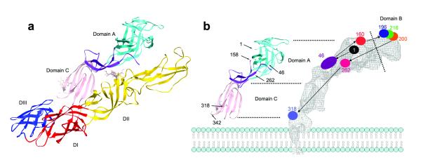
Alphaviruses are enveloped RNA viruses that have a diameter of about 700 Å and can be lethal human pathogens. Entry of virus into host cells by endocytosis is controlled by two envelope glycoproteins, E1 and E2. The E2-E1 heterodimers form 80 trimeric spikes on the icosahedral virus surface, 60 with quasi-three-fold symmetry and 20 coincident with the icosahedral three-fold axes arranged with T = 4 quasi-symmetry. The E1 glycoprotein has a hydrophobic fusion loop at one end and is responsible for membrane fusion. The E2 protein is responsible for receptor binding and protects the fusion loop at neutral pH. The lower pH in the endosome induces the virions to undergo an irreversible conformational change in which E2 and E1 dissociate and E1 forms homotrimers, triggering fusion of the viral membrane with the endosomal membrane and then releasing the viral genome into the cytoplasm. Here we report the structure of an alphavirus spike, crystallized at low pH, representing an intermediate in the fusion process and clarifying the maturation process. The trimer of E2-E1 in the crystal structure is similar to the spikes in the neutral pH virus except that the E2 middle region is disordered, exposing the fusion loop. The amino- and carboxy-terminal domains of E2 each form immunoglobulin-like folds, consistent with the receptor attachment properties of E2.


Figures

Figure 1
Figure 1
The structural proteins of an alphavirus. a, The cryo-EM density of Sindbis virus showing T=4 symmetry. The four E2 molecules in one asymmetric unit (outlined in black) are colored red, green, blue and yellow. These give rise to one trimeric spike on each icosahedral threefold axis and one generally positioned spike. The E1 molecules are colored grey. b, Threading of the Sindbis virus structural polyprotein through an endoplasmic reticulum membrane showing the position of the capsid, E3, E2, 6K and E1 proteins.
Figure 2
Figure 2
Stereo diagrams showing the trimeric spike structure. a, The E1 molecule in a Sindbis virus spike (blue) compared with the E1 molecules in the crystal structure (red) b, linear representation of polypeptides showing domains D-A (cyan), D-B (green), D-C (pink) and the β-ribbon connector (purple) in E2; as well as the domains DI (red), DII (yellow), and DIII (blue) in E1. c, Crystal structure of the trimeric spike at low pH. Domain B is disordered.
Figure 3
Figure 3
The E2-E1 heterodimer. a, The crystal structure (left) color coded as in Fig. 2b. b, Comparison of the earlier E2 mapping with the E2 crystal structure. Amino acid sequence numbers are given in strategic positions. The lipid envelope is shown diagrammatically. [Reprinted from Mukhopadhyay, S. et al. Mapping the structure and function of the E1 and E2 glycoproteins in alphaviruses. Structure 14, 63-73, (2006).]
Figure 4
Figure 4
Cartoon showing maturation and fusion. Color-coded as in Fig. 2b. a, The viral spike while being transported through the Golgi and TGN. Domain B is attached to domain II because E3 (grey) binds the β-ribbon connector to domain A and holds E2 in place at low pH. b, The viral spike on the virus after E3 has been proteolytically cleaved and, in Sindbis virus, has been jettisoned. c, After the virus has recognized a host cell and entered into a low pH endosomal vesicle, domain B becomes unattached from DII, exposing the fusion loop (marked with an *). The dashed line indicates the disordered β-ribbon connector. d, The trimeric spike disassembles, allowing escape of E2 and formation of E1 homotrimers.

Comment in

Similar articles

Cited by

References

    1. Strauss JH, Strauss EG. The alphaviruses: gene expression, replication, and evolution. Microbiol. Rev. 1994;58:491–562. - PMC - PubMed
    1. von Bonsdorff CH, Harrison SC. Sindbis virus glycoproteins form a regular icosahedral surface lattice. J. Virol. 1975;16:141–145. - PMC - PubMed
    1. Kielian M. Membrane fusion and the alphavirus life cycle. Adv. Virus Res. 1995;45:113–151. - PubMed
    1. White J, Helenius A. pH-dependent fusion between the Semliki Forest virus membrane and liposomes. Proc. Natl. Acad. Sci. U.S.A. 1980;77:3273–3277. - PMC - PubMed
    1. Dubuisson J, Rice CM. Sindbis virus attachment: isolation and characterization of mutants with impaired binding to vertebrate cells. J. Virol. 1993;67:3363–3374. - PMC - PubMed

Publication types

MeSH terms